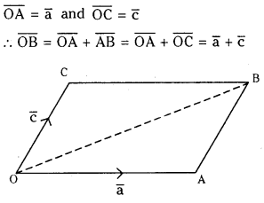
Question 11. Let a̅, b̅ and c̅ be non coplanar vectors. If [2a̅ – b̅ + 3c̅, a̅ + b̅ – 2c̅, a̅ + b̅ – 3c̅] = λ [a̅ b̅ c̅] then find the value of λ.
Answer: Given a,b,c as non coplanar vectors We have [a̅ b̅ c̅] ≠ 0 ∴ ∣∣∣∣211−1113−2−3∣∣∣∣[a̅ b̅ c̅] = [2 (- 3 + 2) + 1 (- 3 + 2) + 3 (1 – 1)][a̅ b̅ c̅] = [-2 – 1] [a̅ b̅ c̅] = -3[a̅ b̅ c̅] Given [2a̅ – b̅ + 3c̅, a̅ + b̅ – 2c̅, a̅ + b̅ – 3c̅] = λ [a b c] We have -3[a̅ b̅ c̅] = λ [a̅ b̅ c̅] ⇒ λ = -3
Question 12. Let a̅, b̅ and c̅ be non coplanar vectors. If [a̅ + 2b̅ 2b̅ + c̅ 5c̅ + a̅] = λ [a̅ b̅ c̅], then find λ.
Answer: Given a̅, b̅ and c̅ as non coplanar vectors. We have [a̅ b̅ c̅] ≠ 0. Given that ∴ ∣∣∣∣101220015∣∣∣∣[a̅ b̅ c̅] = λ[a̅ b̅ c̅] ⇒ [1 (10 – 0) – 2 (0 – 1)] [a̅ b̅ c̅] = λ[a̅ b̅ c̅] ⇒ 12 [a̅ b̅ c̅] = λ [a̅ b̅ c̅] ⇒ λ = 12
Question 13. If a̅, b̅, c̅ are non coplanar vectors, then find the value of (a¯¯¯+2b¯¯¯−c¯¯¯)⋅[(a¯¯¯−b¯¯¯)×(a¯¯¯−b¯¯¯−c¯¯¯)][a¯¯¯b¯¯¯c¯¯¯]
Answer: Given a̅, b̅, c̅ are non coplanar we have [a̅ b̅ c̅] ≠ 0, then = 1 (1) – 2 (- 1) – 1 (- 1 + 1) = 3
Question 14. If a̅, b̅, c̅ are mutually perpendicular unit vectors, then find the value of [a̅ b̅ c̅]2.
Answer: Given a̅, b̅, c̅ are mutually perpendicular unit vectors. We have |a̅| = |b̅| = |c̅| = 1 and taking a̅ = i̅, b̅ = j̅, c̅ = k̅ We have [a̅ b̅ c̅] = [i̅ j̅ k̅] = i̅ . (j̅ × k̅) = i̅.i̅ = 1 ∴ [a̅ b̅ c̅]2 = 1
Question 15. a̅, b̅, c̅ are non zero vectors and a̅ is perpendicular to both b̅ and c̅. If |a̅|= 2, |b̅|= 3, |c̅| = 4 and (b̅, c̅) = 2π3 then find |[a̅ b̅ c̅]|. (May 2008)
Answer: Given a̅, b̅, c̅ are non zero vectors and a is perpendicular to both b̅ and c̅ ⇒ a̅ is parallel to (b̅ × c̅) ⇒ (a̅, b̅ × c̅) = 0 (or) 180° ∴ [a̅ b̅ c̅] = [a̅ . b̅ × c̅]
Question 16. If a̅, b̅, c̅ are unit coplanar vectors, then find [2a̅ – b̅ 2b̅ – c̅ 2c̅ – a̅].
II. Question 1. If [b̅ c̅ d̅] + [c̅ a̅ d̅] + [a̅ b̅ d̅] = [a̅ b̅ c̅], then show that the points with position vectors a̅, b̅, c̅ and d̅ are coplanar. (May 2014)
Question 2. If a̅, b̅ and c̅ are non coplanar vectors, then prove that the four points with position vectors 2a̅ + 3b̅ – c̅, a̅ – 2b̅ + 3c̅, 3a̅ + 4b̅ – 2c̅ and a̅ – 6b̅ + 6c̅ are coplanar.
Answer: Suppose A, B, C, D are the given points with respect to a fixed origin ‘O’ and given that OA¯¯¯¯¯¯¯¯ = 2a̅ + 3b̅ – c̅, OB¯¯¯¯¯¯¯ = a̅ – 2b̅ + 3c̅ OC¯¯¯¯¯¯¯¯ = 3a̅ + 4b̅ – 2c̅ and OD¯¯¯¯¯¯¯¯ = a̅ – 6b̅ + 6c̅ ∴ AB¯¯¯¯¯¯¯=OB¯¯¯¯¯¯¯−OA¯¯¯¯¯¯¯¯ = (a̅ – 2b̅ + 3c̅) – (2a̅ + 3b̅ – c̅) = -a̅ – 5b̅ + 4c̅
Question 3. a̅, b̅ and c̅ are non zero and non collinear vectors and θ ≠ 0, is the angle between b̅ and c̅. If (a̅ × b̅) × c̅ = 13|b̅||c̅||a̅| find sin θ.
Answer: Given |a̅| ≠ 0, |b̅| ≠ 0, |c̅| ≠ 0 and (b̅, c̅) = θ and (a̅ × b̅) × c̅ = 13|b̅||c̅|a̅ ⇒ (a̅ . c̅) b̅ – (b̅ . c̅) a̅ = 13|b̅||c̅|a̅ ∵ a,b, c are non collinear vectors
Question 4. Find the volume of the tatrahedron whose vertices are (1, 2, 1), (3, 2, 5), (2. – 1, 0) and (- 1, 0, 1). (Mar. 2015-T.S) [May 2007]
Answer: Let O be the origin with A, B, C, D as vertices of tetrahedron.
Question 5. Show that (a̅ + b̅) . (b̅ + c̅) × (c̅ + a̅) = 2 [a̅ b̅ c̅]
Question 6. Show that the equation of the plane passing through the points with position vectors 3i̅ – 5j̅ – k̅, -i̅ + 5j̅ + k̅ and parallel to the vector 3i̅ – j̅ + 7k̅ is 3x + 2y – z = 0.
Answer: Let OA¯¯¯¯¯¯¯¯ = (3i̅ – 5 j̅ – k̅), OB¯¯¯¯¯¯¯ = – i̅ + 5j̅ + 7k̅ The given plane passes through the points A, B and parallel to the vector OC¯¯¯¯¯¯¯¯ = 3i̅ – j̅ + 7k̅, AB¯¯¯¯¯¯¯=OB¯¯¯¯¯¯¯−OA¯¯¯¯¯¯¯¯ = -4i̅ + 10j̅ + 8k̅ ∴ Equation of the plane is = x (70 + 8) – y (- 28 – 24) + z (4 – 30) = 78x + 52y – 26z = 26 (3x + 2y – z)
Question 8. If a̅, b̅, c̅ and d̅ are coplanar vectors, then show that (a̅ × b̅) × (c̅ × d̅) = 0.
Answer: Given a̅, b̅, c̅, d̅ are coplanar vectors ⇒ a̅ × b̅ is perpendicular to the plane S. In the similar way c̅ × d̅ is perpendicular to the plane S. a̅ × b̅ and c̅ × d̅ are parallel vectors. ⇒ (a̅ × b̅) × (c̅ × d̅) = 0 (or) (a̅ × b̅) × (c̅ × d̅) = [a̅ c̅ d̅]b̅ – [b̅ c̅ d̅]a̅ = 0b̅ – 0a̅ = 0 (∵ a̅, b̅, c̅, d̅ are coplanar)
Question 9. Show that [(a̅ × b̅) × (a̅ × c̅)] d̅ = (a̅ . d̅) [a̅ b̅ c̅]
Question 12. Find the vector equation of the plane passing through the intersection of planes. r̅ -(2i̅ + 2j̅ – 3k̅) = 7, r̅ = (2i̅ + 5j̅ + 3k̅) = 9 and through the point (2, 1, 3).
Answer: The planes are of the form r̅.n̅1 = d1 and r̅.n̅2 = d2 The vector equation of the plane passing through the intersection of above plane is of the form r̅ . (n̅1 + λn̅2) = d1 + λd2 ∴ r̅ . [(2i̅ + 2j̅ – 3k̅) + λ(2i̅ + 5j̅ + 3k̅)] = 7 + 9λ
Denote r̅ = xi̅ + yj̅ + zk̅ ………..(1) then (2x + 2y – 3z) + λ (2x + 5y + 3z)] = 7 + 9λ ⇒ (2x + 2y – 3z – 7) + λ (2x + 5y + 3z – 9) = 0 …..(2) Since this plane passes through the point (2, 1, 3) We have (4 + 2 – 9 – 7) + λ (4 + 5 + 9 – 9) = 0 ⇒ – 10 + 9λ = 0 ⇒ λ = 109 From (2), (2 + 2λ) x + (2 + 5λ) y + (3λ – 3) z – (7 + 9λ) = 0
Question 13. Find the equation of the plane passing through (a̅, b̅, c̅) and parallel to the plane r̅. (i̅ + j̅ + k̅) = 2.
Answer: Given equation of the plane is r̅ . (i̅ + j̅ + k̅) = 2 Suppose r̅ = xi̅ + yj̅ + zk̅ then (xi̅ + yj̅ + zk̅) . (i̅ + j̅ + k̅) = 2 ⇒ x + y + z = 2 Equation of parallel plane is x + y + z = k Since this passes through (a, b, c) we have a + b + c = k Equation of the required plane is x + y + z = a + b + c
Question 14. Find the shortest distance between the lines r̅ = 6i̅ + 2j̅ + 2k̅ + λ(i̅ – 2j̅ + 2k̅) and r̅ = -4i̅ – k̅ + μ(3i̅ – 2j̅ – 2k̅).
Answer: Given lines are Shortest distance between the lines = 9 units.
Question 15. Find the equation of the plane passing through the line of intersection of the planes r̅.(i̅ + j̅ + k̅) = l and r̅.(2i̅ + 3j̅ – k̅) + 4 = 0 and parallel to X – axis.
Answer: Cartesian form of the given plane is x + y + z = 1 and 2x + 3y – z + 4 = 0 Equation of required plane will be of the form (x + y + z – 1) + λ (2x + 3y – z + 4) = 0 …………(i) ⇒ (1 + 2λ)x + (1 + 3λ)y + (1 – λ)z – (1 – 4λ) = 0 Since this is parallel to X-axis coefficient of x = 0 ⇒ 1 + 2λ = 0 ⇒ λ = 12 Required plane equation from (1) is (x + y + z – 1) –12(2x + 3y – z + 4) = 0 ⇒ 2x + 2y + 2z – 2 – 2x – 3y + z – 4 = 0 ⇒ y – 3z + 6 = 0
Question 16. Prove that the four points 4i̅ + 5j̅ + k̅, -(j̅ + k̅), 3i̅ + 9j̅ + 4k̅ and -4i̅ + 4j̅ + 4k̅ are coplanar.
Question 17. If a̅, b̅, c̅ are non coplanar, then show that the vectors a̅ – b̅, b̅ + c̅, c̅ + a̅ are coplanar.
Answer: Given that a̅, b̅, c̅ are non coplanar we have [a̅ b̅ c̅] ^O ∴ [a̅ – b̅ b̅ + c̅ c̅ + a̅] = ∣∣∣∣101−110011∣∣∣∣[a̅ b̅ c̅] = [1 + 1 (-1)][a̅ b̅ c̅] = 0 [a̅ b̅ c̅] = 0 ∴ Vectors a̅ – b̅, b̅ + c̅, c̅ + a̅ are coplanar.
Question 18. If a̅, b̅, c̅ are the position vectors of the points A, B and C respectively, then prove that the vector a̅ × b̅ + b̅ × c̅ + c̅ × a̅ is perpendicular to the plane of ΔABC.
Answer: Let O be the origin and = (b̅ × c̅) – (b̅ × a̅) – (a̅ × c̅) + (a̅ × a̅) = (b̅ × c̅) + (a̅ × b̅) + (c̅ × a̅) (∵ (a̅ × a̅) = 0) = (a̅ × b̅) + (b̅ × c̅) + (c̅ × a̅) Hence (a̅ × b̅) + (b̅ × c̅) + (c̅ × a̅) is perpendicular to the plane of ΔABC.
An intimate association between two organisms of different species in which, one is benefited and the other one is often adversely affected is called parasitism. The organism that obtains nourishment is called parasite and the organism from which the nourishment is obtained is called host.
Question 2.
Distinguish between facultative parasite and obligatory parasite.
Answer:
a) Facultative Parasite : They lead parasitic life on host if available or may lead free living life in its absence. Example : (Mycobacterium tuberculosus), Ascaris lumbricoides is facultative anaerobe. b) Obligatory parasite : They lead total parasitic life on host and in its absence, they die. Example : (Blood fluke) Taenia solium is an obligatory anaerobe.
Question 3.
Distinguish between definitive host and intermediate host.
Answer:
Definitive host : It is the host that harbours the adult stage or sexually mature stage of a parasite or the host in which the parasite undergoes sexual reproduction. Intermediate host : It is the host that harbours the developing larval or immature or asexual stages of a parasite or the host in which the parasite undergoes asexual reproduction.
Question 4.
Distinguish between vector and a reservoir host.
Answer:
a) Reservoir host : It is the host that lodges the infective stages of a parasite in its body when the main host is not available. In the reservoir host, the parasite neither undergoes development nor causes any disease. b) Vector : It is an organism which transfers the infective stages of a parasite from one main host to another.
Question 5.
Distinguish between mechanical vector and biological vector.
Answer:
a) Mechanical vector : It is the vector, which merely transfers the infective stages of a parasite but no part of the parasitic development takes place in it. b) Biological vector : It is the vector in which the parasite undergoes a part of the development before it gets transferred to another host.
Question 6.
What is a hyper-parasite? Mention the name of one hyper-parasite.
Answer:
Hyperparasite : It is a parasite which lives in /on the body of another parasite. eg : Nosema notabilis (cnidosporan parasite) lives in Sphaerospora polymorpha (also a cnidosporan parasite) which lives in the urinary bladder of toad fish.
Question 7.
What do you mean by parasitic castration? Give one example. [March 2020, 17]
Answer:
Parasitic castration : Some parasites cause the degeneration of gonads of the host, making it sterile. This effect is called parasitic castration, eg : Sacculina (a crustacean) causes degeneration of ovaries in the crab carcinus.
Question 8.
What are the parasitic adaptations observed in Ascaris lumbricoides?
Answer:
1.Developed protective cuticle to withstand the action of digestive enzymes of the host. 2.Reproductive potential is high due to risky lifecycle. Ascaris female lays 2 lakhs of eggs per day. 3.It is a facultative anaerobe.
Question 9.
What are the endo-parasitic adaptations observed in Fasciola hepatica?
Answer:
Life cycle of Fasciola hepatica is very complex involving many developmental stages and two intermediate hosts to increase the chances of reaching a new definitive host.
Question 10.
Define neoplasia. Give one example.
Answer:
Neoplasia : Some cause an abnormal growth of the host cells in a tissue to form . new structures. This effect is called Neoplasia which leads to cancers, eg : Some viruses.
Question 11.
Define the most accurate definition of the term ‘health’ and write any two factors that affect the health.
Answer:
Health is a state of complete physical, mental and social well-being and not merely ‘absence of any disease’ or ‘absence of physical fitness’. Two factors that affect the health are 1) contaminated water 2) pollution of air.
Question 12.
Distinguish between infectious and non-infectious diseases. Give two examples each.
Answer:
a) Infectious diseases : The diseases which are easily transmitted from one person to another are called infectious diseases. Eg. Amoebic dysentery, Malaria, b) Non – infectious diseases : The diseases which are not transmitted from one person to another and are not caused by pathogens are called non-infectious diseases. eg : Kidney problems, genetic disorders, heart problems.
Question 13.
When can you diagnose a healthy person as unhealthy?
Answer:
When the functioning of one or more organs or systems of the body is adversely affected characterized by various signs and symptoms, we say that we are not healthy or we are unhealthy.
Question 14.
Write any two diagnostic features of trophozoite of Entamoeba histolytica.
Answer:
a) Cartwheel shaped nucleus. b) Single blunt finger like pseudopodium called lobopodium. c) Presence of RBC in food vacuole.
Question 15.
‘Entamoeba histolytica is an obligatory anaerobe.’ Justify.
Answer:
The absence of mitochondria indicates the obligate anaerobic nature of Entamoeba histolytica.
Question 16.
Distinguish between precystic stage and cystic stage of E. histolytica.
Answer:
Precystic stage : It is the non-feeding and non-pathogenic stage of Entamoeba histolytica. It is a small, spherical or oval, non-motile form. Cystic stage : It is round in shape and is surrounded by a thin, delicate, and highly resistant cyst wall. This is infective stage.
Question 17.
What is the reserve food in the precystic and early cyst stages of Entamoeba histolytica?
Answer:
The reserve food in the precystic and early cyst stages of Entamoeba histolytica are glycogen granules and chromatoid bars (made up of ribonucleo protein).
Question 18.
What is a metacystic form with reference to Entamoeba histolytica?
Answer:
When the tetranucleate cysts of entamoeba histolytica enter a new human host, they pass into small intestine where the cyst wall gets ruptured by the action of the enzyme trypsin, releasing the tetranucleate amoebae. Such tetranucleate excystic amoebae are called metacysts.
Question 19.
A person is suffering from bowel irregularity, abdominal pain, blood and mucus in stool /etc. Based on these symptoms, name the disease and its causative organism.
Answer:
The disease is Amoebic dysentery or Amoebiasis. The causative organism is Entamoeba histolytica.
Question 20.
On the advice of a doctor, a patient has gone to a clinical laboratory for the examination of a sample of faeces. The lab technician, on observing the stool of the patient diagnosed that the patient was suffering from amoebiasis. Write any two characteristic features based on which the technician came to that conclusion.
Answer:
1.Stools with blood and mucous. 2.Stools containing the tetranucleate cysts.
Question 21.
Define ‘asymptomatic cyst passers’ with reference to Entamoeba histolytica.
Answer:
Some people do not exhibit any symptoms of amoebic dysentery or intestinal amoebiasis or tropical amoebiasis. Such people are called carriers or ‘asymptomatic cyst passers’ as their stool contains the tetranucleate cysts.
Question 22.
Distinguish between primary amoebiasis and secondary amoebiasis.
Answer:
1.Stools with blood and mucous with acute abdominal pair is generally referred to as primary amoebiasis. 2.When trophozoites rupture the wall of capillaries, enter the blood stream, and primarily reach the liver where they may cause abscesses – some call it secondary amoebiasis.
Question 23.
What are the stages of Plasmodium vivax that infect the hepatocytes of man?
Answer:
The stages of plasmodium vivax that infect the hepatocytes of man are 1.Sporozoites primarily 2.Cryptozoites Secondarily (They are also referred to as I generation merozoites)
Question 24.
What are the stages of Plasmodium vivax that infect the RBC of the intermediate host?
Answer:
The stages of Plasmodium vivax that infect the RBc of the intermediate host are a) Cryptozoites b) Micro-metacryptozoites.
Question 25.
Define prepatent period. What is its duration in the life cycle of Plasmodium vivax?
Answer:
The interval between the first entry of plasmodium into the blood in the form of sporozoites and the second entry of plasmodium into the blood in the form of cryptozoites is called “Prepatent period”. In Plasmodium vivax it is 8 days.
Question 26.
Define incubation period. What is its duration in the life cycle of Plasmodium vivax? [March 2018 – A.P.]
Answer:
The period between the entry of plasmodium into the blood in the form of sporozoites and the first appearance of symptoms of malaria in man is called incubation period. It is approximately 10 to 14 days in Plasmodium vivax.
Question 27.
What are Schuffner’s dots? What is their significance?
Answer:
In the signet ring stage’of malarial parasite small red coloured dots appear in the cytoplasm of the RBC known as Schuffner’s dots. These are believed to be the antigens released by the parasite.
Question 28.
What are haemozoin granules ? What is their significance?
Answer:
The malaria parasite digests the globin part of the ingested haemoglobin and converts the soluble haem into an insoluble crystalline haemozoin granules. It is called the malaria pigment which is a disposable product.
Question 29.
Distinguish between schizogony and sporogony.
Answer:
1.In man the plasmodium reproduces by asexual reproduction called Schizogony. It occurs in liver cells as well as in RBC. 2.The formation of sporozoites in the oocysts seen in the stomach wall of female Anopheles mosquito is called SPOROGONY.
Question 30.
What is exflagellation and what are the resultant products called?
Answer:
The formation of male gametes or microgametes by lashing movements like flagella from micro gametocyte is called exflagellation.
Question 31.
Why is the syngamy found in Plasmodium called anisogamy?
Answer:
In plasmodium the male and female gametes are dissimilar in size, the process of union or syngamy is called Anisogamy.
Question 32.
What is ookinete? Based on the ‘sets of chromosomes’ how do you describe it?
Answer:
The zygote remains inactive for some time and then transforms into a long, slender, motile, vermiform ookinete or vermicule within 18 – 24 hours. The ookinete is diploid.
Question 33.
What is tertian fever, with reference to the types of malaria you have learnt about? Give the name of the causative species of the pathogen concerned.
Answer:
Four species of Plasmodium cause four types of malaria in man. They are a) Plasmodium vivax – benign tertian malaria. b) Plasmodium falciparum – malignant tertian malaria or cerebral malaria. c) Plasmodium ovale – mild tertian malaria. d) Plasmodium malariae-quartan malaria. Recurrent fever of Malaria is called tertian fever.
Question 34.
What is the significance of hypnozoites, with reference to malaria fever?
Answer:
Some of the stages of macro-metacryptozoites may survive for a long period in liver as dormant stages called HYPNOZOITES. Reactivation of these hypnozoites leads to the initiation of fresh erythrocytic cycles resulting in the new attacks of malaria. This is referred to as relapse of malaria.
Question 35.
A person is suffering from chills and shivering and high temperature. These symptoms arp cyclically followed by profuse sweating and return to normal body temperature. Based on these symptoms, name the disease and its causative organism.
Answer:
a) Malaria is the disease; b) Causative organism Plasmodium.
Question 36.
Describe the methods of biological control of mosquitoes.
Answer:
Biological control of mosquitoes : Introduction of larvivorous fishes like Gambusia, insectivorous plants like utricularia into the places where mosquitoes breed.
Question 37.
The eggs of Ascaris are called ‘mammillated eggs’. Justify.
Answer:
In Ascaris each egg is surrounded by a protein coat with rippled surface. Hence the eggs of Ascaris are described as “mammillated eggs”.
Question 38.
Write the route of extra intestinal migration followed by the juveniles of Ascaris lumbricoides.
Answer:
Extra intestinal migration exhibited by 2nd stage larva of Ascaris.
Question 39.
Write any two differences between male and female worms of Wuchereria bancrofti.
Answer:
Wuchereria bancrofti male and female differences. a) Male worm : Its posterior end is curved with a cloacal aperture. A pair of unequal, chitinous pineal spicules or copulatory spicules is present in the cloacal region. b) Female worm : Its posterior end is straight. Anus is present near the posterior end. The female genital pore or vulva is present mid ventrally at about one third the length from the mouth. It is ovi viviparous.
Question 40.
What is meant by nocturnal periodicity with reference to the life history of a nematode parasite you have studied?
Answer:
The microfilaria larvae of Wuchereria bancrofti in man move in the peripheral blood circulation during the night time between 10 pm and 4 am. This tendency is referred to as NOCTURNAL PERIODICITY.
Question 41.
Distinguish between lymphadenitis and lymphangitis.
Answer:
1.Inflammation in the lymph vessels is called lymphangitis. 2.Inflammation in the lymph glands is called lymphadenitis. Both are caused by filarial worm injection.
Question 42.
‘Elephantiasis is the terminal condition of filariasis.’ Justify.
Answer:
Fibroblasts accumulate in these tissues and form the fibrous tissue. In severe cases, the sweat glands of the skin in the affected regions disintegrate and the skin becomes rough. This terminal condition is referred to as Elephantiasis.
Question 43.
Mention the pathogens that cause ringworm.
Answer:
Ringworm is one of the most common infectious diseases in man. It is caused by many fungi belonging to the genera Microsporum, Trichophyton and Epidermophyton.
Question 44.
Explain any three preventive measures to control microbial infections.
Answer:
1.The immunization programmes by the use of vaccines have enabled us to completely eradicate a deadly disease like smallpox. 2.Biotechnology is at the verge of making available newer and safer vaccines. 3.Discovery of antibiotics and various other drugs has also enabled us to treat infectious diseases effectively.
Question 45.
“Maintenance of personal and public hygiene is necessary for prevention and control of many infectious diseases.” Justify the statement giving suitable examples.
Answer:
The following hygienic habits help prevent spread of this disease. a) Using boiled and filtered water b) Washing hands, fruits and vegetables properly c) Using septic tank toilets These habits prevent diseases like Amoebiasis and Ascariasis.
Question 46.
Diseases like amoebic dysentery, ascariasis, typhoid etc., are more common in overcrowded human settlements. Why?
Answer:
In overcrowded human settlements, poor sanitation facilities are seen. No septic toilets for defecation. Drinking water is only from available water sources like tanks, lakes and ponds where cleaning of cattle, washing of clothes is also done. Due to these conditions the contaminated water, food and air spread the diseases like typhoid, amoebic dysentery and ascariasis.
Question 47.
In which way does tobacco affect the respiration? Name the alkaloid found in tobacco : [May 2017 – A.P.]
Answer:
Tobacco is smoked or chewed as gutkha or used in the form of snuff. Smoking increases the carbon monoxide level and reduces the oxygen level in the blood. Alkaloid found in tobacco is NICOTINE.
Question 48.
Define drug abuse.
Answer:
When drugs are taken for a purpose other than the medicinal use or in excess amounts that impair one’s physical or psychological functions, it constitutes “drug abuse”.
Question 49.
From which substances ‘Smack’ and ‘Coke’ are obtained?
Answer:
Smack and coke are obtained from : Smack is chemically diacetylmorphine obtained by the acetylation of morphine extracted from dried latex of unripe seed capsule of poppy plant. Coke or crack is obtained from coca plant Erythroxylum coca, commonly called cocaine.
Question 50.
‘Many secondary metabolites of plants have medicinal properties. It is their misuse that creates problems.’ Justify the statement with an example.
Answer:
Benzodiazepines (tranquilizers), Lysergic acid diethyl amides (LSD) and other similar drugs, normally used as medicines to treat patients with mental illnesses like depression are often abused.
Question 51.
Write the scientific names of any two plants with hallucinogenic properties.
Answer:
Question 52.
Why are cannabinoids and anabolic steroids banned in sports and games?
Answer:
Cannabinoids and anabolic steroids (AAS) increase protein synthesis within cells which result in the build up of cellular tissues, especially in muscles. Cannabinoids show their effects on cardio-vascular system of the body. Hence they are banned in sports and games (DOPING TEST). They give tremendous energy temporarily.
Question 53.
Mention the names of any four drugs which are used as medicines to treat patients with mental illness like depression, insomnia, etc.’ that are often abused.
What is the need for parasites to develop special adaptations? Mention some special adaptations developed by the parasites. [May 2017 – A.P.]
Answer:
Parasites have evolved special adaptations to meet the requirements and lead successful life in the hosts. 1.In order to live in the host, some parasites have developed structures like hooks, suckers, rostellum, etc., for anchoring, e.g. Taenia solium. 2.Some intestinal parasites have developed protective cuticle to withstand the action of the digestive enzymes of the host. e.g. Ascaris lumbricoides. 3.Some intestinal parasites produce anti enzymes to neutralize the effect of host’s digestive enzymes, e.g. Taenia solium. 4.Some parasites live as obligatory anaerobes as the availability of oxygen is very rare for them. e.g. Entamoeba histolytica, Taenia solium, etc. 5.Some intestinal parasites live as facultative anaerobes i.e., if oxygen is not available, they live anaerobically and if oxygen is available, they respire aerobically, e.g. Ascaris lumbricoides. 6.The morphological and anatomical features are greatly simplified while emphasizing their reproductive potential. For example, an Ascaris lays nearly two lakh eggs per day. In Taenia solium the body is divided into 700 to 900 proglottids of which each proglottid acts as a unit of reproductive system and releases approximately 35,000 eggs. 7.The life cycles of endoparasites are more complex because of their extreme specialization. For example, life cycle of certain parasites like Fasciola hepatica (sheep liver fluke) is very complex involving many developmental stages and two intermediate hosts to increase the chances of reaching a new definitive host. 8.Certain parasites like Entamoeba develop cysts to tide over the unfavourable conditions like desiccation while reaching the new host. 9.Some parasites elude production of vaccines against them (smart parasites!) as they keep changing their surface antigens form time to time. e.g. Plasmodium, HIV, etc.
Question 2.
Describe the effects of parasites on the host.
Answer:
Effects of parasites on hosts : In general, the parasites cause weakening of the body of their hosts by causing the deprivation of nutrients, fluids and metabolites as they compete with their hosts for the same. They may also cause pathological effects in their hosts suchas 1) Parasitic castration : Some parasites cause the degeneration of gonads of the host, making it sterile. This effect is called parasitic castration. e.g. Sacculina (root headed barnacle, a crustacean) causes the degeneration of ovaries in the crab Carcinus maenas. 2) Neoplasia : Some cause an abnormal growth of the host cells in a tissue to form new structures. This effect is called neoplasia which leads to cancers, e.g. Some viruses 3) Gigantism : Some parasites cause an abnormal increase in the size of the host. This effect is called gigantism, e.g. The larval stages of Fasciola hepatica cause gigantism in snail (an intermediate host). 4) Hyperplasia : Some parasites cause an increase in the number of cells. This effect is called hyperplasia, e.g. Fasciola hepatica in the bile ducts of sheep. 5) Hypertrophy : Some parasites cause an abnormal increase in the volume / size of the infected host cells. This effect is called hypertrophy, e.g. RBC of man infected by Plasmodium. 6) Most of the parasites cause various types of diseases like 1.African sleeping sickness by Trypanosoma gambiense 2.Delhi boils / Tashkent ulcers / Oriental sores by Leishmania tropica 3.Kala azar/ Dum dum fever/Visceral leishmaniasis by Leishmania donovani 4.Malaria by Plasmodium sps 5.Elephantiasis by Wuchereria bancrofti.
Question 3.
Distinguish between hypertrophy and hyperplasia with an example [March 2020]
Answer:
Hyperplasia : Some parasites cause an increase in the number of cells. This effect is called hyperplasia, e.g. Fasaola hepatica in the bile ducts of sheep Hypertrophy: Some parasites cause an abnormal increase in the volume / size of the infected host cells. This effect is called hypertrophy, e.g. RBC of man infected by Plasmodium
Question 4.
Write any four types of parasitic diseases. Mention the primary and secondary hosts of these parasites.
Answer:
1.Malaria caused by Plasmodium. Primary host – female Anopheles mosquito. Secondary host-man. 2.Elephantiasis caused by wuchereria bancrofti. Primary host – man. Secondary host – female culex mosquito. 3.Amoebic dysentery or Anoebiasis caused by Entamoeba histolytica. Single host -man. 4.Ascariasis caused by Ascaris lumbricoides. Single host – man.
Question 5.
Describe the structure of a trophozoite of Entamoeba histolytica.
Answer:
Trophozoite stage : It is the most active, motile, feeding and pathogenic stage that lives in the mucosa and sub-mucosa membranes of the large intestine. It moves with the help of a single blunt finger like pseudopodium called lobopodium which is produced anteriorly. The body of the trophozoite is surrounded by plas- malemma. Its cytoplasm is differentiated into an outer clear, viscous, non-granular ectoplasm and the inner fluid like, granular endoplasm. Ribosomes, food vacuoles and a vesicular, cartwheel shaped nucleus are present in the endoplasm. However contractile vacuoles, endoplasmic reticulum, Golgi apparatus and mitochondria are absent. The absence of mitochondria indicates the ‘obligate anaerobic nature’ of Entamoeba histolytica. It produces the proteolytic enzyme called histolysin due to which the species name histolytica’ was assigned to it. Due to the effect of this enzyme, the mucosa and sub-mucosa of the gut wall are dissolved releasing some amount of blood, tissue debris which are ingested by the trophozoites. Hence, the food vacuoles are with erythrocytes, fragments of epithelial cells and bacteria. The mode of nutrition is holozoic. Presence of ‘RBC in food vacuoles’ and cartwheel shaped nucleus are the characteristic features of the trophozoites of Entamoeba histolytica.
Question 6.
Explain the life cycle of Entamoeba histolytica.
Answer:
Life cycle : The trophozoites undergo binary fissions in the wall of the large intestine and produce a number of daughter entamoebae. They feed upon the bacteria and the host’s tissue elements, grow in size and again multiply. After repeated binary fissions, when the trophozoites increase in number, some of the young ones enter the lumen of the large intestine and transform into precystic stages. Here, the precystic stages transform into cystic stages which in turn develop into tetranucleate cysts. The entire process is completed only in a few hours. These tetranucleate cysts come out along with the faecal matter and can remain alive for about 10 days. These cysts reach new host through contaminated food and water. They pass into the small intestine of a new human host where the cyst wall gets ruptured by the action of the enzyme trypsin, releasing the tetranucleate amoebae. Such tetranucleate excystic amoebae are called metacysts. The four nuclei of the metacyst undergo mitotic divisions and produce eight nuclei. Each nucleus gets a bit of the cytoplasm and thus eight daughter entamoebae or ‘metacystic trophozoites’ are produced. These young ones develop into feeding stages called trophozoites. They invade the mucous membrane of the large intestine and grow into mature trophozoites.
Question 7.
Write a short note on the pathogenicity of Entamoeba histolytica.
Answer:
The trophozoites ‘dissolve’ the mucosal lining by histolysin, go deep into sub¬mucosa and cause ulcers. These ulcers contain cellular debris, lymphocytes, blood corpuscles and bacteria. It leads to the formation of abscesses in the wall of large intestine. Ultimately it results in stool with blood and mucous. This condition is called amoebic dysentery or intestinal amoebiasisor tropical amoebiasis. Some people do not exhibit any symptoms. Such people are called ‘carriers or asymptomatic cyst passers’ as their stool contains the tetranucleate cysts. They help in spreading the parasites to other persons. Extra-intestinal amoebiasis: Sometimes, the trophozoites may rupture the wall of capillaries, enter the blood stream and primarily reach the liver where they may cause ‘abscesses’ (some call it ‘secondary amoebiasis’). From there, they may go to lungs, heart, brain, kidneys, gonads, etc., and cause abscesses in those parts leading to severe pathological conditions.
Question 8.
Describe the structure of sporozoite of Plasmodium vivax.
Answer:
Structure of sporozoite : It is sickle shaped with a swollen middle part and pointed at both ends of its body. It measures about 15 microns in length and one micron in width. The body is covered by an elastic pellicle with microtubules which help in the wriggling movements of the sporozoite. The cytoplasm contains cell organelles such as Golgi complex, endoplasmic reticulum, mitochondria and a nucleus. Cytoplasm also shows many convoluted tubules of unknown function throughout the length of the body. It contains a cup like depression called apical cup at the anterior end into which a pair of secretory organelles opens. They secrete a cytolytic enzyme, which helps in the penetration of sporozoite into the liver cells.
Question 9.
What do you know about the exo-erythrocytic cycle of plasmodium vivax?
Answer:
This is a part of the life cycle of Plasmodium taking place in the liver of man. This starts after the liberation of cryptozoites from ruptured liver cells. Exo-erythrocytic cycle: If the cryptozoites enter the fresh liver cells, they undergo the changes similar to that of the pre-erythrocytic cycle and produce the second generation merozoites called metacryptozoites. These are of two types – the smaller micro-metacryptozoites and larger macro-metacryptozoites. This entire process is completed approximately in two days. The macro-metacryptozoites attack fresh liver cells and continue another exo-erythrocytic cycle, whereas the micro- metacryptozoites always enter blood stream and attack fresh RBC to continue erythrocytic cycle.
Question 10.
Describe the cycle of Golgi in the life history of Plasmodium vivax.
Answer:
Erythrocytic cycle : Itwas first described by Camillo Golgi. Hence it is also called Golgi cycle. This cycle is initiated either by the cryptozoites of pre-erythrocytic cycle or the micro-metacryptozoites of exo-erythrocytic cycle. In the fresh RBC, these stages assume spherical shape and transform into trophozoites. It develops a small vacuole which gradually enlarges in size, pushing the cytoplasm and nucleus to the periphery. Now the Plasmodium looks like a finger ring. Hence this stage is called signet ring stage. Soon it loses the vacuole, develops pseudopodia and becomes amoeboid stage. With the help of pseudopodia, it actively feeds on the contents of the RBC and increases in size. As a result, the RBC grows almost double the size, this process is called hypertrophy. The malaria parasite digests the globin part of the ingested haemoglobin and converts the soluble haem into an insoluble crystalline haemozoin. It is called the ‘malaria pigment’ which is a disposable product. During this stage, small red coloured dots appear in the cytoplasm of the RBC known as Schuffner s dots. These are believed to be the antigens released by the parasite. Now the Plasmodium loses the pseudopodia, further increases in size, occupies the entire RBC and becomes a schizont. It undergoes schizogony similar to that of the pre-erythrocytic cycle and produces 12 to 24 erythrocytic merozoites. They are arranged in the form of the petals of a rose in the RBC. Hence, this stage is called the rosette stage. Finally the erythrocyte bursts and releases the merozoites along with haemozoin into the blood. This cycle is completed approximately in 48 hours.
Question 11.
Describe the process of sporogony in the life cycle of Plasmodium vivax. What is the significance of sporozoites?
Answer:
Sporogony : The formation of sporozoites in the oocysts is called sporogony. According to Bano, the nucleus of the oocyst first undergoes reduction division followed by repeated mitotic divisions resulting in the formation of about 1,000 daughter nuclei. Each bit of nucleus is surrounded by a little bit of the cytoplasm and transforms into a sickle shaped sporozoite. Oocyst with such sporozoites is called sporocyst. When this sporocyst ruptures, the sporozoites are liberated into the haemocoel of the mosquito. From there, they travel into the salivary glands and are ready for infection. The life cycle of Plasmodium in mosquito is completed in about 10 to 24 days.
Question 12.
Explain the pathogenicity of Wuchereria bancrofti in man.
Answer:
Pathogenicity of Wuchereria bancrofti in man : Light infection causes filarial fever which is characterised by headache, mental depression and increase in the body temperature. In general, the infection of filarial worm causes inflammatory effect in lymph vessels and lymph glands. Inflammation in the lymph vessels is called lymphangitis and that of lymph glands is called lymphadenitis. In the case of heavy infection, the accumulation of dead worms blocks the lymph vessels and lymph glands resulting in immense swelling. This condition is called lymphoedema which is noticed in the extremities of limbs, scrotum of males and mammary glands in females. Fibroblasts accumulate in these tissues and form the fibrous tissue. In severe cases, the sweat glands of the skin in the affected regions disintegrate and the skin becomes rough. This terminal condition is referred to as elephantiasis.
Question 13.
Write short notes on typhoid fever and its prophylaxis.
Answer:
Typhoid fever : It is caused by Salmonella typhi which is a Gram negative bacterium. It mainly lives in the small intestine of man and then migrates to other organs through blood. It can be confirmed by Widal test. Mode of infection : Contamination through food and water. Symptoms : Sustained fever with high temperature upto 104 F, weakness, stomach pain, constipation, headache and loss of appetite. Intestinal perforation and death may also occur in severe cases. Prophylaxis : The immunization programmes by the use of vaccines have enabled us to completely eradicate a deadly disease like smallpox. Biotechnology is at the verge of making available newer and safer vaccines. Discovery of antibiotics and various other drugs has also enabled us to treat infectious diseases effectively.
Question 14.
Write short notes on pneumonia and its prophylaxis.
Answer:
Pneumonia : It is caused by Gram positive bacteria such as streptococcus pneumoniae and Haemophilus influenzae. They infect the alveoli of lungs in human beings. Mode of infection : Contamination by inhaling the droplets / aerosols released by an infected person or even by sharing the utensils with an infected person. Symptoms : The alveoli get filled with fluid leading to severe problems in respiration. In severe cases, the lips and finger nails may turn gray to bluish in colour. Prophylaxis : The immunization programmes by the use of vaccines have enabled us to completely eradicate a deadly disease like smallpox. Biotechnology is at the verge of making available newer and safer vaccines. Discovery of antibiotics and various other drugs has also enabled us to treat infectious diseases effectively.
Question 15.
Write short notes on common cold and its prophylaxis.
Answer:
Common cold : It is caused by Rhino virus group of viruses. They infect nose and respiratory passage but not lungs. Mode of infection : Contamination by direct inhalation of the droplets resulting from cough or sneezes of an infected person or indirectly through contaminated objects such as pens, books, cups, door-knobs, computer keyboard or mouse etc. Generally all the medicines that are used against cold cause drowsiness. Symptoms : Nasal congestion, discharge from nose, sore throat, hoarseness, cough, headache, tiredness, etc., which usually last for 3 – 7 days. Prophylaxis : The immunization programmes by the use of vaccines have enabled us to completely eradicate a deadly disease like smallpox. Biotechnology is at the verge of making available newer and safer vaccines. Discovery of antibiotics and various other drugs has also enabled us to treat infectious diseases effectively.
Question 16.
Write short notes on ‘ringworm’ and its prophylaxis.
Answer:
Ringworm : It is one of the most common infectious diseases in man. It is caused by many fungi belonging to the genera Microsporum, Trichophyton, and Epidermophyton. Heat and moisture help these fungi grow in the skin folds such as those in the groin or between the toes. Mode of infection : Contamination by using towels, clothes or combs of the infected persons or even from soil. Symptoms : Appearance of dry, scaly, usually round lesions accompanied by intense itching on various parts of the body such as skin, nails and scalp. Prophylaxis : Using of antifungal drugs like Griesoven orally, and antifungal creams like Zole, Itchguard. Ring guard for external tise’cures the disease. Hygiene habits prevent the disease.
Question 17.
What are the adverse effects of tobacco?
Answer:
Tobacco has been used by human beings for more than 400 years. It contains a large number of chemical substances including nicotine, an alkaloid. While buying cigarettes one cannot miss the statutory warning present on the packet Smoking is injurious to health. Mode of abuse : It is smoked or chewed as gutkha or used in the form of snuff. Effect : Smoking increases the carbon monixide (CO) level and reduces the oxygen level in the blood. Nicotine stimulates the adrenal gland to release adrenaline and nor-adrenaline into blood. These hormones raise the blood pressure and increase the heart rate. Smoking is associated with bronchitis, emphysema, coronary heart disease, gastric ulcer and increases the incidence of cancers of throat, lungs, urinary bladder etc. Smoking also paves the way to hard drugs. Yet, smoking is very prevalent in society, both among young and old. Tobacco chewing is associated with increased risk of cancer of the oral cavity.
Question 18.
Write short notes on opioids.
Answer:
Opioids : These are the drugs obtained from opium poppy plant Papaver somniferum (vernacular name : Nallamandu mokka). They bind to specific opioid receptors present in our central nervous system and gastrointestinal tract. Some of them are morphine, heroin, etc. i) Morphine : It is extracted from the dried latex of the unripe seed capsule (pod) of poppy plant. It occurs as colourless crystals or a white crystalline powder. Mode of abuse : Generally it is taken orally or by injection. Effect : It is a very effective sedative and painkiller. It is very useful in patients who have undergone surgery. ii) Heroin : It is a white, bitter, odourless and crystalline compound, obtained by the acetylation of morphine. Chemically it is diacetylmorphine. It is commonly called ‘smack’. Mode of abuse : Generally it is taken by snorting and injection. Effect : Heroin is a depressant and slows down the body functions.
Question 19.
Write short notes on Cannabinoids.
Answer:
Cannabinoids : These are a group of chemicals obtained from Indian hemp plant Cannabis sativa (vernacular name: Ganjai mokka). They interact with cannabinoid receptors present in the brain. The flower tops, leaves and the resin of this plant are used in various combinations to produce marijuana, hashish, charas and ganja. These days, cannabinoids are being abused by even some sports-persons (doping). Mode of abuse : These are generally taken by inhalation and oral ingestion. Effect : Show their effects on cardiovascular system of the body.
Question 20.
Write short notes on Cocaine.
Answer:
Coca alkaloid or Cocaine : It is a white, crystalline alkaloid that is obtained from the leaves of Coca plant Erythroxylum coca, native to South America. It is commonly called coke or crack. Mode of abuse : It is usually snorted. Effect : It has a potent stimulating action on the central nervous system as it interferes with the transport of the neuro transmitter dopamine. Hence it produces a sense of euphoria and increased energy. Its excessive dosage causes hallucinations. Other well-known plants with hallucinogenic properties are Atropa belladonna and Datura. Certain drugs like ‘Barbiturates (sleeping pills), Amphetamines (cause sleeplessness), Benzodiazepines (tranquilizers), Lysergic acid diethyl amides (LSD) and other similar drugs, normally used as medicines to treat patients with mental illnesses like depression, insomnia, etc.,1 are often abused.
Question 21.
Why is adolescence considered vulnerable phase?
Answer:
Adolescence : It is the time period between the beginning of puberty and the beginning of adulthood. In other words, it is the bridge linking childhood and adulthood. The age between 12 -18 years is considered adolescence period. It is both ‘a period and a process’ during which a child becomes mature. It is accompanied by several biological and behavioural changes. Thus, adolescence is a very vulnerable phase of mental and psychological development of an individual.
Question 22.
Why do some adolescents start taking drugs? How can this be avoided?
Answer:
Curiosity, desire for adventure and excitement, experimentation, are the common causes for the motivation of youngsters towards the use of tobacco, drugs. The first use of drugs or alcohol may be out of curiosity or experimentation, but later the person starts using them to escape facing problems. Recently ‘stress from the pressure to excel in academics or examination’s has played a significant role in alluring the youngsters to try certain drugs. Television, movies, newspapers and internet also help promoting this wrong perception. Other factors that are associated with tobacco, drug and alcohol abuse among adolescents are unstable or unsupportive family structures and peer pressure. A lot of help is available in the form of highly qualified psychologists, psychiatrists and de-addiction and rehabilitation programmers. Educating and counselling the adolescents to face problems, stress and failures as a part of life.
Question 23.
Distinguish between addiction and dependence. [March 2017 -A.P.]
Answer:
Addiction : It is a psychological attachment to certain effects such as euphoria. The most important thing one fails to realise is, the inherent addictive nature of tobacco, drugs and alcohol. With the repeated use of TDA, the tolerance level of the receptors present in our body increases. Consequently the receptors respond only to higher doses leading to greater intake and addiction. However it should be clearly borne in mind that use of TDA even once, can be a fore-runner to addiction. Thus, the addictive potential of tobacco, drugs’and alcohol pull the users into a vicious circle leading to their regular use (abuse) from which they may not be able to get out. In the absence of any guidance or counseling, people get addicted and become dependent on them. Dependence : It is the tendency of the body to manifest a characteristic and unpleasant condition (withdrawal syndrome) if the regular dose of drugs or alcohol is abruptly discontinued. The withdrawal syndrome is characterised by anxiety, shakiness (tremors), nausea and sweating which may be relieved when the regular use is resumed again. Dependence leads the patients to ignore all social norms.
Question 24.
“Prevention is better than cure.” Justify with regard to TDA abuse. [March 2018 – A.P.]
Answer:
Prevention and Control: The age-old adage of Prevention is better than cure holds true here also. Some of the measures useful for prevention and control of TDA abuse among the adolescents are : i) Avoid undue parental pressure : Every child has his / her own choice, capacity and personality. The parents should not force their children to perform beyond their capacity by comparing them with others in studies, games, etc. ii) Responsibility of parents and teachers : They should look for the danger signs and counsel such students who are likely to get into the ‘trap’. iii) Seeking help from peers : If peers find some one abusing drugs or alcohol, immediately it should be brought to the notice of their parents or teachers so that they can guide them appropriately. iv) Education and counselling : Educating and counselling the children to face problems, stress and failures as a part of life. v) Seeking professional and medical help : A lot of help is available in the form of highly qualified psychologists, psychiatrists and de-addiction and rehabilitation programmers.
Essay Answer Type Questions
Question 1.
Explain the structure and life cycle of Entamoeba histolytica with the help of neat and labelled diagrams.
Answer:
Structure: Entamoeba histolytica passes through three distinct stages in its life cycle, namely: 1.Trophozoite stage 2.Precystic stage and 3.Cystic stage
Trophozoite stage : It is the most active, motile, feeding and pathogenic stage that lives in the mucosa and sub-mucosa membranes of the large intestine. It moves with the help of a single blunt finger like pseudopodium called lobopodium which is produced anteriorly. The body of the trophozoite is surrounded by plasmalemma. Its cytoplasm is differentiated into an outer clear, viscous, non-granular ectoplasm and the inner fluid like, granular endoplasm. Ribosomes, food vacuoles and a vesicular, cartwheel shaped nucleus are present in the endoplasm. However contractile vacuoles, endoplasmic reticulum, Golgi apparatus and mitochondria are absent. The absence of mitochondria indicates the ‘obligate anaerobic nature’ of Entamoeba histolytica. It produces the proteolytic enzyme called histolysin due to which the species name histolytica’ was assigned to it. Due to the effect of this enzyme, the mucosa and sub-mucosa of the gut wall are dissolved releasing some amount of blood, tissue debris which are ingested by the trophozoites. Hence, the food vacuoles are with erythrocytes, fragments of epithelial cells and bacteria. The mode of nutrition is holozoic. Presence of ‘RBC in food vacuoles’ and cartwheel shaped nucleus are the characteristic features of the trophozoites of Entamoeba histolytica. Precysticstage :
It is the non-feeding and non-pathogenic stage of Entamoeba histolytica that is found in the lumen of the large intestine. It is a small, spherical or oval, non- motile form. The cytoplasm of the precystic stage stores glycogen granules and chromatoid bars (made of ribonucleo protein) which act as reserve food. Cystic stage :
It is round in shape and is surrounded by a thin, delicate and highly resistant cyst wall. It is found in the lumen of the large intestine. The process of development of cyst wall is called encystation which is a means to tide over the unfavourable conditions that the parasite is going to encounter while passing to a new host. Soon after the encystation, the nucleus undergoes two successive mitotic divisions to form four daughter nuclei. This type of cystic stage is called tetra nucleate cyst or mature cyst which is ‘the stage infective to man’. Life cycle : The trophozoites undergo binary fissions in the wall of the large intestine and produce a number of daughter entamoebae. They feed upon the bacteria and the host’s tissue elements, grow in size and again multiply. After repeated binary fissions, when the trophozoites increase in number, some of the young ones enter the lumen of the large intestine and transform into precystic stages. Here, the precystic stages transform into cystic stages which in turn develop into the tetranucleate cysts. The entire process is compelted only in a few hours. These tetranucleate cysts come out along with the faecal matter and can remain alive for about 10 days. These cysts reach new host through contaminated food and water. They pass into the small intestine of a new human host where the cyst wall gets ruptured by the action of the enzyme trypsin, releasing the tetranucleate amoebae. Such tetranucleate excystic amoebae are called metacysts. The four nuclei of the metacyst undergo mitotic divisions and produce eight nuclei. Each nucleus gets a bit of the cytoplasm and thus eight daughter entamoebae or ‘metacystic trophozoites’ are produced. These young ones develop into feeding stages called trophozoites. They invade the mucous membrane of the large intestine and grow into mature trophozoites. Pathogenicity : The trophozoites ‘dissolve’ the mucosal lining by histolysin, go deep into sub¬mucosa and cause ulcers. These ulcers contain cellular debris, lymphocytes, blood corpuscles and bacteria. It leads to the formatjon of abscesses in the wall of large intestine . Ultimately it results in stool with blood and mucous. This condition is called amoebic dysentery or intestinal amoebiasis or tropical amoebiasis. Some people do not exhibit any symptoms. Such people are called ‘carriers or asymptomatic cyst passers’ as their stool contains the tetranucleate cysts. They help in spreading the parasites to other persons. Extra-intestinal amoebiasis : Sometimes, the trophozoites may rupture the wall of capillaries, enter the blood stream, and primarily reach the liver where they may cause ‘abscesses’ (some call it ‘secondary amoebiasis’). From there, they may go to lungs, heart, brain, kidneys, gonads, etc., cause abscesses in those parts leading to severe pathological conditions.
Question 2.
Describe the life cycle of Plasmodium vivax in man. [March 2017 – A.P.]
Answer:
Life cycle of Plasmodium in man (The human phase): In man, the Plasmodium reproduces by asexual reproduction called schizogony. It occurs in liver cells (hepatocytes) as well as in RBC. In liver cells, it is called hepatic schizogony and in RBC it is called erythrocytic schizogony. Hepatic schizogony : This was discovered by Shortt and Garnham. Whenever, a mosquito infected by Plasmodium bites a man, nearly 2000 sporozoites are released into the blood of man through its saliva. Within half an hour, they reach the hepatocytes where they undergo pre-erythrocytic and exo-erythrocytic cycles. Pre-erythrocytic cycle : Whenever the sporozoites reach the liver cells, they transform into trophozoites. They feed on the contents of the hepatic cells, assume spherical shape and attain the maximum size. This stage is called schizont stage. Its nucleus divides several times mitotically, followed by the cytoplasmic divisions resulting in approximately 12,000 daughter individuals called cryptozoites or the regeneration merozoites. They enter the sinusoids of the liver by rupturing the cell membrane of the schizont and the liver cells. This entire process is completed approximately in 8 days. Now these first generation merozoites have two options i.e., they can enter either fresh liver cells and continue exo-erythrocytic cycle or they can enter RBC and continue erythrocytic cycle. Exo-erythrocytic cycle : If the cryptozoites enter the fresh liver cells, they undergo the changes similar to that of the pre-erythrocytic cycle and produce the second generation merozoites called metacryptozoites. These are of two types -the smaller micro-metacryptozoites and larger macro-metacryptozoites. This entire process is completed approximately in two days. The macro-metacryptozoites attack fresh liver cells and continue another exo – erythrocytic cycle, whereas the micro- metacryptozoites always enter blood stream and attack fresh RBC to continue erythrocytic cycle. Prepatent period : The interval between ‘the first entry of Plasmodium into the blood in the form of sporozoites and the second entry of Plasmodium into the blood in the form of cryptozoites is called prepatent period. It lasts approximately 8 days. During this period, the host does not show any clinical symptoms of the disease. It is only a means of multiplication. Erythrocytic cycle : It was first described by Camillo Golgi. Hence it is also called Golgi cycle. This cycle is initiated either by the cryptozoites of pre-erythrocytic cycle or the micro-metacryptozoites of exo – erythrocytic cycle. In the fresh RBC, these stages assume spherical shape and transform into trophozoites. It develops a small vacuole which gradually enlarges in size, pushing the cytoplasm and nucleus to the periphery. Now the Plasmodium looks like a finger ring. Hence this stage is-” called signet ring stage. Soon it loses the vacuole, develops pseudopodia and becomes amoeboid stage. With the help of pseudopodia, it actively feeds on the contents of the RBC and increases in size. As a result, the RBC grows almost double the size. This process is called hypertrophy. The malaria parasite digests the globin part of the ingested haemoglobin and converts the soluble haem into an insoluble crystalline haemozoin. It is called the ‘malaria pigment’ which is a disposable product. During this stage, small red coloured dots appear in the cytoplasm of the RBC known as Schuffner s dots. These are believed to be the antigens released by the parasite. Now the Plasmodium loses the pseudopodia, further increases in size, occupies the entire RBC and becomes a schizont. It undergoes schizogony similar to that of the pre-erythrocytic cycle and produces 12 to 24 erythrocytic merozoites. They are arranged in the form of the petals of a rose in the RBC. Hence, this stage is called the rosette stage. Finally the erythrocyte bursts and releases the merozoites along with haemozoin into the blood. This cycle is completed approximately in 48 hours. Incubation Period : The period between ‘the entry of Plasmodium into the blood in the form of sporozoite and the first appearance of symptoms of malaria in man’ is called incubation period. It is approximately 10 to 14 days. Formation of gametocytes : After repeated cycles of erythrocytic schizogony, when the number of fresh RBC decreases, some merozoites enter the RBC and transform into gametocytes instead of continuing the erythrocytic cycle. This process generally takes place when the RBCs are present in spleen and bone marrow. The gametocytes are of two types namely, smaller microgametocytes or male gametocytes and larger macrogametocytes or female gametocytes. The gametocytes cannot undergo further development in man as the temperature and pH of the blood of man are not suitable for further development. These gametocytes reach the blood circulation and wait to reach the next host. They degenerate and die if they are not transferred to mosquito within a week.
Question 3.
Describe the life cycle of Plasmodium vivax in mosquito.
Answer:
Life cycle of Plasmodium in mosquito (The mosquito phase)- Ross cycle: When a female Anopheles mosquito bites and sucks the blood of a malaria patient, the gametocytes along with the other stages of the erythrocytic cycle reach the crop of mosquito. Here all the stages are digested except the gametocytes. Further part of the life cycle consists of: 1.Gametogony 2.Fertilization 3.Formation of Ookinete & Oocysts 4.Sporogony i) Gametogony : The formation of male and female gametes from the gametocytes is called gametogony. It occurs in the lumen of the crop of mosquito. Formation of male gametes : During this process, the nucleus of microgametocyte divides into eight daughter nuclei called pronuclei which reach the periphery. The cytoplasm is pushed out in the form of eight flagella like processes. Into each flagellum like process, one pronucleus enters and forms a micro gamete or male gamete. These male gametes show lashing movements like flagella and get separated from the cytoplasm of microgametocyte. This process is called exflagellation. Formation of female gamete : The female gametocyte undergoes a few changes and transforms into a female gamete. This process is called maturation. The nucleus of the female gamete moves towards the periphery and the cytoplasm at that point forms a projection. This projected region is called the fertilization cone. ii) Fertilization : The fusion of male and female gametes is called fertilization. It also occurs in the lumen of the crop of the mosquito. When an actively moving male gamete comes into contact with the fertilization cone of the female gamete, it enters it. The pronuclei and cytoplasm of these two gametes fuse with each other, resulting in the formation of a synkaryon. Since the two gametes are dissimilar in size, this process is known as anisogamy. The female gamete that bears the synkaryon is called the zygote which is round and non – motile.
iii) Formation of ookinete and oocysts : The zygote remains inactive for some time and then transforms into a long, slender, motile, vermiform ookinete or vermicide within 18 to 24 hours. It pierces the wall of the crop and settles beneath the basement membrane. It becomes round and secretes a cyst around its body. This encysted ookinete is now called oocyst. About 50 to 500 oocysts are formed on the wall of the crop and appear in the form of small nodules. (Sir Ronald Ross identified these oocysts for the first time). iv) Sporogony :
The formation of sporozoites in the oocysts is called sporogony. According to Bano, the nucleus of the oocyst first undergoes reduction division followed by repeated mitotic divisions resulting in the formation of about 1,000 daughter nuclei. Each bit of nucleus is surrounded by a little bit of the cytoplasm and transforms into a sickle shaped sporozoite. Oocyst with such sporozoites is called sporocyst. When this sporocyst ruptures, the sporozoites are liberated into the haemocoel of the mosquito. From there, they travel into the salivary glands and are ready for infection. The life cycle of Plasmodium in mosquito is completed in about 10 to 24 days.
Question 4.
Describe the structure and life cycle of Ascaris lumbricoides with the help of a neat and labelled diagram.
Answer:
Structure : Sexes are separate and the sexual dimorphism is distinct. In both males and females, the body is elongated and cylindrical. Mouth is present at the extreme anterior end and is surrounded by three chitinous lips. Close to the mouth mid ventrally, there is a small aperture called excretory pore. Male : It has a curved posterior end which is considered the tail. The posterior end possesses a cloacal aperture and a pair of equal sized chitinous ‘pineal spicules or ‘pineal setae which serve to transfer the sperms during copulation. Female : It has a straight posterior end, the tail. The female genital pore or vulva is present mid ventrally at about one third the length from mouth. The anus is present a little in front of the tail end. Life history : Copulation takes place in the small intestine of man. After copulation, the female releases approximately two lakh eggs per day. Each egg is surrounded by ‘a protein coat with rippled surface. Hence the eggs of Ascaris are described as mammillated eggs. The protein coat is followed by a chitinous shell and a lipid layer internally. These eggs come out along with faecal matter. In the moist soil, development takes place inside the egg so that the 1st stage rhabditiform larva is produced. It undergoes the 1st moulting and becomes the 2nd stage rhabditiform larva which is considered ‘the stage infective to man’. They reach the alimentary canal of man through contaminated food and water.
In the small intestine, the shell gets dissolved so that the 2nd stage larva is released. Now it undergoes extra intestinal migration. First it reaches the liver through the hepatic portal vein. From there it reaches the heart through the post caval vein. It goes to the lungs through the pulmonary arteries. In the alveoli of lungs it undergoes the 2nd moulting to produce the 3rd stage larva. It undergoes the 3rd moulting so that the 4th stage larva is produced in the alveoli only. It leaves the alveoli and reaches the small intestine again, through bronchi, trachea, larynx, glottis, pharynx, oesophagus and stomach. In the small intestine, it undergoes the 4th and final moulting to become a young one which attains sexual maturity within 8 to 10 weeks. Pathogenicity : The disease caused by Ascaris lumbricoides is called ascariasis. The disease is asymptomatic if the number of worms is less. A heavy infection causes nutritional deficiency and severe abdominal pain. It also causes stunted growth in children.
Question 5.
Describe the life cycle of Wuchereria bancrofti.
Answer:
Life Cycle : Wuchereria bancrofti completes its life cycle in two hosts namely man and female culex mosquito. In man : Both male and female worms are found coiled together in the lymphatic vessels of man. After copulation the female releases the sheathed microfilaria larvae into the lymph of man. Each sheathed microfilaria larva measures 0.2 to 0.3 mm in length. It is surrounded by a loose cuticular sheath which is supposed to be the modified shell. They migrate to the blood circulation and reside in the deeper blood vessels during the day time. They move to the peripheral blood circulation during the night time between 10.00 pm and 4.00 am. This tendency is referred to as nocturnal periodicity. When a female Culex mosquito sucks the blood of an infected person, they enter the gut of mosquito. They die if they are not transferred to mosquito with in 70 days. In mosquito : In the mid gut of mosquito, the sheath of the larva is dissolved within 2 to 6 hours of the infection. The ex-sheathed microfilaria larva penetrates the gut wall and reaches the haernocoel of mosquito. From there, it reaches the thoracic muscles and transforms into a ‘sausage shaped larva within two days. It is called the first stage larva or first stage microfilaria. This undergoes two moultings within 10 to 20 days and transforms into a long, infective 3rd stage microfilaria. It reaches the labium of the mosquito.
In man after the infection : When an infected mosquito bites a man, the 3rd stage microfilaria larvae enter the blood circulation of man and finally reach the lymphatic vessels. Here they undergo the 3rdand the 4th moultings to produce young filarial worms. They attain sexual maturity within 5 to 18 months.
Question 6.
Write an essay on adolescence and TDA abuse.
Answer:
Adolescence and TDA abuse: Adolescence : It is the time period between the beginning of puberty and the beginning of adulthood. In other words, it is the bridge linking childhood and adulthood. The age between 12 -18 years is considered adolescence period. It is both ‘a period and a process’ during which a child becomes mature. It is accompanied by several biological and behavioural changes. Thus, adolescence is a very vulnerable phase of mental and psychological development of an individual. TDA abuse : Curiosity, desire for adventure and excitement, experimentation, are the common causes for the motivation of youngsters towards the use of tobacco, drugs and alcohol. The first use of drugs or alcohol may be out of curiosity or experimentation, but later the person starts using them to escape facing problems. Recently ‘stress from the pressure to excel in academics or examinations’ has played a significant role in alluring the youngsters to try certain drugs. Television, movies, newspapers and internet also help promoting this wrong perception. Other factors that are associated with tobacco, drug and alcohol abuse among adolescents are unstable or unsupportive family structures and peer pressure. Addiction and Dependence : The TDA abuse leads to addiction and dependence. Addiction : It is a psychological attachment to certain effects such as euphoria. The most important thing one fails to realise is, the inherent addictive nature of tobacco, drugs and alcohol. With the repeated use of TDA, the tolerance level of the receptors present in our body increases. Consequently the receptors respond only to higher doses leading to greater intake and addiction. However it should be clearly borne in mind that use of TDA even once, can be a fore-runner to addiction. Thus, the addictive potential of tobacco, drugs and alcohol pull the users into a vicious circle leading to their regular use (abuse) from which they may not be able to get out. In the absence of any guidance or counselling, people get addicted and become dependent on them. Dependence : It is the tendency of the body to manifest a characteristic and unpleasant condition (withdrawal syndrome) if the regular dose of drugs or alcohol is abruptly discontinued. The withdrawal syndrome is characterised by anxiety, shakiness (tremors), nausea and sweating which may be relieved when the regular use is resumed again. Dependence leads the patients to ignore all social norms. Adverse effects of drugs and alcohol abuse : The immediate adverse effects of drugs and alcohol abuse are manifested in the form of reluctant behaviour, vandalism and violence. Excessive doses of drugs may lead to coma and death due to respiratory or heart failure or cerebral haemorrhage. A combination of drugs or their intake along with alcohol generally results in overdosing and even death. The most common warning signs of drug and alcohol abuse among the youth include drop in academic performance, lack of interest in personal hygiene, depression, fatigue, aggressive behaviour, loss of interest in hobbies, change in sleeping and eating habits, fluctuations in weight, appetite, etc. Those who take drugs intravenously are much more likely to acquire serious infections such as HIV, HBV (Hepatitis – B virus) etc., as the viruses are transferred from one person to another by the sharing of infected needles and syringes. The chronic use of drugs and alcohol damages nervous system and liver. The use of drugs and alcohol during pregnancy is also known to affect the foetus adversely. Some sports-persorTs take drugs such as anabolic steroids to enhance their performance. The side-effects of the use of these drugs in females include masculinisation, increased aggressiveness, mood swings, depression, abnormal menstrual cycles, excessive hair growth on the face and body and the enlargement of clitoris. In males it includes acne (pimples), increased aggressiveness, mood swings, depression, reduction in the size of testicles, decreased sperm production, kidney and liver dysfunction, enlargement of breasts, premature baldness and the enlargement of the prostate gland. Prevention and Control : The age-old adage of Prevention is better than cure holds true here also. Some of the measures useful for prevention and control of TDA abuse among the adolescents are : i) Avoid undue parental pressure : Every child has his / her own choice, capacity and personality. The parents should not force their children to perform beyond their capacity by comparing them with others in studies, games, etc. ii) Responsibility of parents and teachers : They should look for the danger signs and counsel such students who are likely to get into the ‘trap’. iii) Seeking help from peers : If peers find someone abusing drugs or alcohol, immediately it should be brought to the notice of their parents or teachers so that they can guide them appropriately. iv) Education and counselling : Educating and counselling the children to face problems, stress and failures as a part of life. v) Seeking professional and medical help: A lot of help is available in the form of highly qualified psychologists, psychiatrists and de-addiction and rehabilitation programmers.
Draw a labelled diagram of T.S. of flagellum. [March 2018-A.P.]
Answer:
Question 2.
List any two differences between a flagellum and a cilium. [Mar. 2020, ’19, 17 – A.P ; May/June, Mar. 2014]
Answer:
Flagellum
Cilium
1) The long whip-like locomotor organelles are called flagella. Found in Mastigophoran Protozoans
1) These are small hair-like structures found in ciliate protists, genital ducts, respiratory tract.
2) Flagellum helps only in locomotion.
2) Cilia serve as organelles or locomotion, food collection and also act as Sensory structures.
3) Flagella perform undular movement.
3) Cilia perform pendular movement.
Question 3.
What are dynein arms? What is their significance? [May 2017 – A.P.]
Answer:
In cross section of flagellum / cilium Dynein arms are seen. The ‘A’ tubule of flagellum/ cilium of each peripheral doublet bears paired arms along its length, called dynein arms Bending movement of a flagellum is brought about by the sliding of microtubules past each other due to the functioning of ‘dynein arms’.
Question 4.
What is a Kinety? [March 2014]
Answer:
A longitudinal row of kinetosomes together with Kinetodesmata constitute a unit called ‘Kinety’.
Question 5.
Distinguish between synchronous and metachronous movements.
Answer:
1) Synchronous movement : Cilia in a transverse row beat simultaneously in one direction. It is called synchronous movement. 2) Metachronous movement : The sequential movement of Cilia, in a longitudinal row, one after the other in one direction is called metachronous movement.
Question 6.
Why do we refer to the offspring, formed by asexual method of reproduction, a clone? [March 2020, 19]
Answer:
The term clone is used to describe morphologically and genetically similar daughter individuals formed from single parent by asexual reproduction. They are not only identical to one another but also exact copies of their parent.
Question 7.
Distinguish between proter and opisthe. [Mar. 2017, 13 – A.P ; Mar. 2015 A.P & T.S]
Answer:
During Transverse binary fission of paramecium, two daughter paramecia are formed. The upper or anterior one is proter which receive upper contractile vacuole, cytopharynx and cytostome of its parent. The lower or posterior daughter is opisthe which receives the posterior contractile vacuole only.
Question 8.
How is sexual reproduction advantageous in evolution?
Answer:
Because of the fusion of male and female gametes, sexual reproduction results in offspring that are not identical to the parents or amongst themselves. This leads gradually to origin of new species in course of evolution.
Question 9.
Distinguish between lobopodium and filopodium. Give an example to each of them. [May ’17 ; March 2015 – A.P.]
Answer:
a) Blunt finger like pseudopodia are called lobopodium and are seen in Amoeba and Entamoeba. b) Fibre like pseudopodia are called filopodiurn and are seen in Euglypha.
Question 10.
Define conjugation with reference to ciliates. Give two examples. [March 2018 – A.P.; March 2015 – T.S ; May/June 2014] A. Conjugation is a temporary union between two senile ciliates that belong to two different mating types for the exchange of nuclear material and its reorganization. This is observed in Paramecium and Vorticella.
Short Answer Type Questions
Question 1.
Name the system that controls the fastest swimming movement of protozoans and write its components.
Answer:
The system that controls the fastest swimming movement of protozoans is infraciliary system. It is located just below the pellicle in the ectoplasm of a ciliate. It includes kinetosomes, kinetodesmal fibrils and kinetodesmata. The kinetosomes are present at the bases of cilia in transverse and longitudinal rows. The kinetodesmal fibrils are connected to the kinetosomes and run along the right side of each row of kinetosomes as a ‘cord of fibres’ called kinetodesmata. A longitudinal row of kinetosomes together with kinetodesmata constitute a unit called ‘kinety’. All the kineties together form an infraciliary system, which is connected to a ‘motorium’, located near the cytopharynx. The infraciliary system and motorium form the ‘neuromotor system’ that controls and coordinates the movement of cilia.
Question 2.
Write the mechanism of bending of flagellum and explain effective and recovery strokes.
Answer:
Bending movement of flagella: Dynein arms show a complex cycle of movements using energy provided by ATP (dynein arms are the sites of ATPase activity in the cilia and flagella). The dynein arms of each doublet attach to an adjacent doublet and pull the neighbouring doublet. So the doublets slide past each other in opposite directions. The arms release and reattach a little farther on the adjacent doublet and again ‘puli’. As the doublets of a flagellum or cilium are physically held in place by the radial spokes, the doublets cannot slide past much. Instead they curve and cause bending of flagellum or cilium. Such bending movements of flagella and cilia play an important role in the flagellar and ciliary locomotion.
i) Effective stroke : Flagellum becomes rigid and starts bending to one side beating against the water. This beating against water is at right angles to the body axis and the organism moves forwards. ii) Recovery stroke : Flagellum becomes comparatively soft so as to offer least resistance to water and moves backwards to its original position. It is called ‘recovery stroke’.
Question 3.
What are lateral appendages? Based on their presence and absence, write the various types of flagella giving at least one example for each type. [Mar. 17 – A.P ; Mar. 15 – A.P & T.S]
Answer:
Lateral appendages : Some flagella bear one or two or many rows of short, lateral hair like fibrils called lateral appendages. They are of two types namely ‘mastigonemes’ and ‘flimmers’. Types of Flagella : Based on the presence or absence and / or the number of rows of lateral appendages, five types of flagella are recognised. a. Stichonematic : This flagellum bears one row of lateral appendages on the axoneme. Eg : Euglena and Astasia. b. Pantonematic : This flagellum has two or more rows of lateral appendages on the axoneme. Eg: Peranema and Monas. c. Acronematic : This type of flagellum does not bear lateral appendages and the terminal part of the axoneme is naked without the outer sheath at its tip. Eg: Chlamydomonas and Polytoma.
d. Pantacronematic : This type of flagellum is provided with two or more rows of lateral appendages and the axoneme ends in a terminal naked filament. Eg. Urceolus. e. Anematic or simple : In this type of flagellum, lateral appendages and terminal filament are absent. Hence, it is called anematic (a – no; nematic – threads). Eg: Chilomonas and Cryptomonas.
Question 4.
Describe the process of transverse binary fission in paramecium. [May 2017 – A.P.; May/June 2014]
Answer:
During favourable conditions, Paramecium stops feeding after attaining its maximum growth. At first the micronucleus divides by mitosis and the macronucleus divides into two daughter nuclei by amitosis. The oral groove disappears. After karyokinesis, a transverse constriction appears in the middle of the body, which deepens and divides the parent cell into two daughter individuals, the anterior proter and the posterior opisthe. The proter receives the anterior contractile vacuole, cytopharynx and cytosome from its parent individual. It develops posterior contractile vacuole and a new oral groove. The opisthe receives the posterior contractile vacuole of its parent. It develops a new anterior contractile vacuole, cytopharynx, cytostome and a new oral groove. Binary fission is completed in almost two hours, in favourable conditions and paramecium can produce four generations of daughter individuals by binary fission in a day. The transverse binary fission is also called homothetogenic fission, because the plane of fission is at right angles to the longitudinal axis of the body. As it occurs at right angles to the kineties, it is also called perkinetai fission.
Question 5.
Describe the process of longitudinal binary fission in Euglena. [March 2018 – A.P.; March 2014]
Answer:
During the process of binary fission, the nucleus, basal granules, chromatophores, cytoplasm undergo division. The nucleus divides by mitosis into two daughter nuclei. Then the kinetosomes and the chromatophores also divide. At first, a longitudinal groove develops in the middle of the anterior end. This groove extends gradually towards the posterior end until the two daughter individuals are separated. One daughter Euglena retains the parental flagella. The other daughter individual develops new flagella from the newly formed basal granules. The stigma, paraflagellar body and contractile vacuole of the parent disappear. They develop afresh in both the daughter euglenae. The longitudinal binary fission is known as symmetrogenic division, because the two daughter euglenae resemble each other like ‘mirror images’.
Question 6.
Write a short note on multiple fission.
Answer:
Multiple Fission : It is the division of the parent body into many smaller individuals (Multi – many; Fission – splitting). Normally multiple fission occurs during unfavourable conditions. During the multiple fission, the nucleus first undergoes repeated mitotic divisions without cytokinesis. This causes the formation of many daughter nuclei. Then the cytoplasm also divides into as many-number of bits as there are nuclei. Each cytoplasmic bit encircles one daughter nucleus. This results in the formation of many smaller individuals from a single parent organism. There are different types of multiple fissions in protozoans such as schizogony, male gametogony, sporogony in Plasmodium, sporulation in Amoeba, etc.
Question 7.
Give an account of pseudopodia.
Answer:
Pseudopodia : These are found in rhizopods. The pseudopodia are temporary extensions of cytoplasm that develop in the direction of the movement. These temporary structures are useful to move on the substratum as our legs do, hence the name ‘pseudopodia’. There are four kinds of pseudopodia namely lobopodia (blunt finger-like) as in Amoeba and Entamoeba, filopodia (fibre-like) as in Euglypha, reticulopodia (net-like) as in Elphidium and axopodia or heliopodia (sun ray-like) as in Actionophrys.
The Pseudopodium is formed by the conversion of gel (viscous outer endoplasmic part) to sol (fluid-like inner endoplasmic part) and vice-versa. There are different theories on the formation of pseudopodium. The sol-gel transformation theory is the most accepted theory. Pseudopodial movement is performed by Amoeba, Polystomella, Actionophrys.
Question 8.
Give an account of the ultra structure of an axoneme.
Answer:
The structural components of a typical flagellum include axoneme, microtubules, dynein arms, inner sheath, outer sheath, radial spokes, lateral appendages (such as mastigonemes or flimmers) and a basal granule (kinetosome). i) Axoneme / axial filament : It is the central, longitudinal, microtubular structure of cilium and flagellum. It is surrounded by a membrane which is continuous with plasma membrane. All the components of the axoneme are embedded within the matrix. ii) Microtubules : An axoneme is made up of 2 central ‘singlets’ and 9 peripheral ‘doublets (9+2 array)’. These are formed by the protein, tubulin. Each peripheral doublet consists of an outer ‘A’ (alpha) and the inner ‘B’ (beta) tubules, so, the peripheral tubules are actually 9 microtubular doublets. (The microtubule ‘A’ is smaller but complete whereas ‘B’ is larger and incomplete). The peripheral doublets are interconnected by linkers called nexins. iii) Dynein arms : The ‘A’ tubule of each peripheral doublet bears paired arms along its length, called dynein arms (Dyne – pulling like a dynamo). The dynein arms of ‘A’ tubule face the tubule ‘B’ of the adjacent doublet. They are oriented in the same direction (clockwise) in all microtubules, when the axoneme is viewed from the base to the top. The dynein arms are considered ‘protein motor molecules’. They are made of the protein, dynein. iv) Inner and outer sheaths : The two central singlets are enclosed by a fibrous inner sheath and the peripheral doublets are enclosed by an outer sheath (an extension of plasma membrane). The central singlets do not reach below the level of the pellicle or plasmalemma.
v) Radial spokes : They are elastic fibres that connect the inner sheath, with the ‘A’ tubule of each doublet. They resemble the spokes that connect the rim of a bicycle wheel with the centre, hence the name ‘radial spokes / radial bridges’. The nine radial spokes limit the extent of sliding past of the doublets, during bending movements.
Question 9.
Draw a neat labelled diagram of Euglena.
Answer:
Question 10.
Draw a neat diagram of Paramecium and label its important structures/components.
List out the characters shared by chordates and echinoderms.
Answer:
Chordates share deuterostomeate condition, radial and indeterminate type of cleavage and enterocoelom with the echinoderms.
Question 2.
Write four salient features of cyclostomes.
Answer:
Salient features of cyclostomes 1.These are jawless aquatic forms. 2.Body is scaleless, long, slender and eel-like. 3.Endoskeleton is cartilaginous 4.Mouth is circular and suctorial. Tongue bears horny teeth.
Question 3.
What is the importance of endostyle in lancelets and ascidians?
Answer:
In ascidians and lancelets, ventral side of the pharynx possesses an endostyle which is believed to be the fore runner of the thyroid gland of a vertebrate. It helps in nutrition.
Question 4.
Name the type of caudal fin and scales that are present in a shark and Catla, respectively.
Answer:
1.Shark → Heterocercal caudal fin and placoid scales are seen. 2.Catla → Homocercal caudal fin, ctenoid or cycloid scales are seen.
Question 5.
What is the importance of air bladder in fishes?
Answer:
An air bladder is present with or without connection to the gut. It is either helpful in gas exchange or in maintaining buoyancy.
Question 6.
How do you justify the statement-“heart in fishes is a branchial heart”?
Answer:
Fish heart is known as ‘branchial heart’ as it supplies blood only to the gills.
Question 7.
What are claspers? Which group of fishes possesses them?
Answer:
In the group chondrichthyes males possess claspers on pelvic fins to facilitate internal fertilization, (example : shark).
Question 8.
How does the heart of an amphibian differ from that of a reptile? [March 2015 – T.S.]
Answer:
1.Heart in amphibians is 3 chambered with sinus venosus and conus arteriosus. 2.Heart in reptiles is completely four chambered except in the crocodiles. Sinus venosus is present but conus arteriosus is absent.
Question 9.
Name the structures that appeared for the first time in amphibians, in the course of evolution.
Answer:
Tympanum, Lacrimal and Harderian glands (associated with eye) appeared for the first time in the amphibians in the course of evolution.
Question 10.
How do you distinguish a male frog from a female frog? [March 2018 – A.P.]
Answer:
Male frog can be distinguished by the presence of sound amplifying vocal sacs and also a copulatory pad on the first digit of each forelimb. They are absent in female frogs.
Question 11.
What is ‘force pump’ in frog? Why is it named so?
Answer:
In frog during the pulmonary respiration, the bucco – pharyngeal cavity acts like a “force pump”. Due to the elevation of bucco – pharyngeal cavity the air forces the glottis to open and enter the lungs, where exchange of gases takes place.
Question 12.
What are corporabigemina? Mention their chief function.
Answer:
In amphibians Mid brain is represented by a pair of optic lobes called corpora bigemina. The optic lobes are associated with the sense of sight.
Question 13.
Distinguish between mesorchium and mesovarium.
Answer:
1.In male frog testes are attached to kidney and dorsal body wall by a double fold of peritoneum called mesorchium. 2.In female frog ovaries are attached to the kidneys and dorsal body wall by a double fold of peritoneum called mesovarium.
Question 14.
Distinguish between milt and spawn. [Mar. 2017 – A.P ; May/June 2014,]
Answer:
1.Mass of eggs released by a female frog into water is called Spawn. 2.Mass of sperms released by a male frog into water is called Milt.
Question 15.
What are the “Golden ages” of the first jawed vertebrates and the first amniotes?
Answer:
1.Golden age of first jawed vertebrates is “DEVONIAN PERIOD” (Fishes) 2.Golden age of first amniotes is MESOZOIC ERA (Reptiles)
Question 16.
Name two poisonous and non-poisonous snakes found in South India. [March 2014]
Answer:
1.Two poisonous snakes found in South India are a) Naja naja (cobra), b) Bungarus (Krait) 2.Two non-poisonous snakes found in South India are a) Ptyas (rat snake) b) Tropidonotus (pond snake)
Question 17.
In which features does the skin of a reptile differ from that of a frog?
Answer:
1.Skin of a frog is thin, scaleless and moist. 2.Skin of a reptile is rough and dry. The exoskeleton occurs in the form of horny epidermal scales, shields and claws.
Question 18.
Describe a cat and a lizard on the basis of their chief nitrogenous wastes excreted.
Answer:
1.Reptiles excrete uric acid as nitrogenous waste (uricotelic). 2.Cat excrete urea as nitrogenous waste (ureotelic).
Question 19.
Name the four extra embryonic membranes. [March 2020]
Answer:
The four extra embryonic membranes are a) Amnion b) Allantois c) Chorion and d) Yolk sac.
Question 20.
What are Jacobson’s organs? What is their function?
Answer:
Jacobson’s organs, the specialized olfactory structures are highly developed in lizards and snakes.
Question 21.
What are pneumatic bones? How do they help birds?
Answer:
In birds bones are hollow or air filled and referred to as pneumatic bones. They help in reducing the weight, thus help in effective flying (aerial adaptation).
Question 22.
What is ‘wish bone’? What are the skeletal components that form it?
Answer:
In birds both clavicles are fused with the interclavicle to form a ‘V’- shaped bone, called furcula or ‘wish bone’ or merry thought bone.
Question 23.
What is continuous oxygenation of the blood? How is it made possible in birds?
Answer:
In birds the compact spongy lungs are associated with air sacs. Air sacs facilitate continuous oxygenation (oxygen supplied to lungs uninterruptedly and heavily) of blood and pneumacity of bones.
Question 24.
Distinguish between the crop and the gizzard in birds.
Answer:
a) In birds oesophagus is often dilated into crop for the storage of food. b) Stomach is usually divided into glandular proventriculus and muscular gizzard (grinding mill).
Question 25.
Distinguish between altricial and precocial hatchlings.
Answer:
1.Altricial hatchlings are seen in flying birds which are incapable of moving around on its own soon after hatching. 2.PrecOcial hatchlings are seen in flightless birds which are capable of moving around on its own soon.
Question 26.
In which group of animals do we find three ear ossicles on each side and what are their names from the innermost to the outermost?
Answer:
In mammals, we find three ear ossicles on each side. From innermost to outer¬most they are stapes, incus and malleus.
Question 27.
How does a mature RBC of a mammal differ from that of other vertebrates?
Answer:
Mature RBC in mammals is unique because it is circular, biconcave and enucleate. In other vertebrates RBC are oval, biconvex and nucleated.
Question 28.
Name the characteristic type of vertebrae found in reptiles, birds and mammals.
Name the three meninges. In which group of animals do you find all of them?
Answer:
The three meninges which are brain layers are a) Dura Mater b) Arachnoid membrane c) Piamater. The group mammals possess all the three layers.
Question 30.
Name the vertebrate groups in which ’renal portal system’ is absent.
Answer:
Renal portal system is absent in the group Mammals.
Short Answer Type Questions
Question 1.
Give three major differences between chordstes and non-chordates and draw the sketch of a chordate’s body showing those features.
Answer:
Major differences between chordates and non-chordates:
Chordates
Non-chordates
1. Notochord is present
Notochord is absent
2. Pharynx is perforated by gill slits
Gill slits are absent
3. Heart is ventral
Heart is dorsal (if present).
4. A post-anal tail is present.
Post-anal tail is absent
Question 2.
Name the four ‘hallmarks’ of chordates and explain the principal function of each of them. [March 2020, 18]
Answer:
The four principal chordate characters and their functions are a) Notochord : Flexible rod like structure placed along the mid dorsal line between gut and nerve cord. It is a supporting structure. b) Dorsal tubular nerve cord : A single hollow tubular and fluid filled nerve cord is situated above notochord and below dorsal body wall. It is sensory in function. In higher chordates anteriorly it becomes brain and the rest becomes the spinal cord. c) Pharyngeal gill slits : These are a series of lateral perforations in the wall of pharynx which are ecto-endodermal in origin. They are helpful in the exchange of respiratory gases. d) Post-anal tail : Chordates have a tail extending posterior to the anus. It is lost in many species during embryonic development. It provides the propelling force in the locomotion of aquatic forms and act as a balancing organ in many terrestrial animals.
Question 3.
Describe the features of a tunicate that reveals its chordate identity.
Answer:
SUBPHYLUM – UROCHORDATA OR TUNICATA : All urochordates are marine and occur from the surface water to greater depths. They are either sessile (ascidians) or pelagic (Salpa, Doliolum) and solitary (Ascidia) or colonial (Pyrosoma).
Body is un-segmented and covered by a ‘test’ or ‘tunic’ composed of cellulose1 which is uncommon in animals. Coelom is absent. However, an ectoderm – lined atrial cavity surrounds the pharynx, into which gill slits, anus and genital ducts open. Ventral side of the pharynx possesses an endostyle, (see Glossary) which is believed to be the ‘forerunner’ of the thyroid gland of a vertebrate. Atrium leads to the exterior by a dorsal or posterior atrial aperture. Digestive tract is ‘complete’. Two to numerous gills slits are found in the pharyngeal wall. Circulatory system is of ‘open type’. There is a simple, tubular, ventral heart which alternately reverses the direction of the flow of blood. Nervous system is represented in the adult by a single dorsal ‘ganglion’. They are bisexual or hermaphrodites. Development generally includes a free-swimming tadpole larva with a tail, a dorsal hollow nerve cord, and a notochord confined to the tail, hence the name urochordata. e.g.: Ascidia, Salpa, Doliolum, Pyrosoma and Oikopleura.
Question 4.
Compare and contrast sea squirts and lancelets.
Answer:
Sea squirts
Lancelets
1) Marine and occur from the surface water to greater depths. Sessile or pelagic and solitary or colonial.
1) Marine forms lead a burrowing mode of life in shallow sea waters
2) Body is unsegmented and covered by a test or tunic made up of cellulose
2) Body is fish like, translucent with median fins but without paired fins
3) Larva stage exhibits a tail, dorsal hollow nerve cord and a notochord confined to the tail, hence the name urochordata.
3) Often described as typical chordate because they possess principal chordate. Chordates such as notochord, tubular nerve cord and pharyngeal gill slits throughout their life.
4) Circulatory system is of open type. Simple, tubular, ventral heart which alternately reverses the direction of the flow of blood. Example: Ascidia (sea squirt)
4) Circulatory system is of closed type and heart, blood corpuscles and respiratory pigment are absent. Example: Branchiostoma (lancelet)
Question 5.
List out eight characteristics that help distinguish a fish from the other vertebrates. [March 2019]
Answer:
Characteristics of group PISCES. 1.Fishes are completely aquatic poikilo- thermic (cold blooded ) animals. 2.Body of a fish is usually streamlined and differentiated into head, trunk and tail. 3.Exoskeleton consists of mesodermal scales or bony plates. A few are scaleless. 4.Endoskeleton may be cartilaginous or bony. Skull is monocondylic. Vertebrae are amphicoelous (centrum is concave at both anterior and posterior faces). 5.Locomotion is assisted by unpaired (median and caudal) fins along with paired (pectoral and pelvic) fins. 6.Mouth is ventral or terminal. Teeth are usually acrodont, homodont and polyphyodont. 7.Exchange of respiratory gases is performed by the gills. 8.Heart is ‘two chambered’ and is described as ‘branchial heart’ as it supplies blood only to the gills. 9.Lateral line sensory system is well developed.
Question 6.
Compare and contrast cartilaginous and bony fishes. [May 2017 – A.P.; May/June. Mar. 2014 ; March 2013]
Answer:
Cartilaginous fishes
Bony fishes
1) Caudal fin is heterocercal
1) Caudal fin is diphycercal or homocercal.
2) Scales if present placoid scales.
2) Scales are ganoid, cycloid or ctenoid scales.
3) Endoskeleton is entirely cartilage.
3) Endoskeleton is bony.
4) Mouth and nostrils are ventral
4) Mouth is usually terminal.
5) Digestive tract opens into cloaca, if present.
5) Digestive tract opens out by anus.
6) Air bladder is absent.
6) Air bladder is often present.
7) Fertilization is internal. Mostly viviparous. eg: Scoliodon.
7) Fertilization is external. Mostly oviparous. Eg: Catla catla
Question 7.
Describe the structure of the heart of frog.
Answer:
The heart is a muscular organ situated in the upper part of the body cavity. If has two separate atria and a single undivided ventricle. It is covered bye double layered membrane called pericardium. A triangular chamber called sinus venosus joins the right atrium on the dorsal side. It receives blood through three vena cavae (caval veins). The ventricle opens into the conus arteriosus on the ventral side. The conus arteriosus bifurcates into two branches and each of it divides into three aortic arches namely carotid, systemic and pulmocutaneous. Blood from the heart is distributed to all parts of the body by the branches of aortic arches. Three major veins collect blood from the different parts of the body and carry it to the sinus venosus.
Question 8.
Write eight salient features of the class Amphibia.
Answer:
1.Body is divided into distinct’head’ and’trunk’. Tail may or may not be present. 2.Skin is soft, scale-less (except the members of Apoda), moist and glandular. 3.The body bears two pairs of equal or unequal pentadactyle limbs (caecilians are limbless). 4.Skull is dicondylic as in mammals. Vertebrae are mostly procoelous (centrum is concave at its anterior face only) in the anurans, amphicoelous in the caecilians and usually opisthocoelous (centrum is concave at its posterior face) in the urodeles. Sternum appeared for the first time in the amphibians. 5.Mouth is large; teeth are acrodont, homodont and polyphyodont. 6.Respiratory gaseous exchange is mostly cutaneous; pulmonary and bucco pharyngeal respirations also occur. Branchial respiration is performed by larvae and some adult urodeles. 7.Heart is three-chambered with sinus venosus and conus arteriosus. Three pairs of aortic arches and well-developed portal systems are present; erythrocytes are nucleate. 8.Kidneys are mesonephric; ureotelic. 9.Meninges are the inner piamater and outer duramater; cranial nerves are 10 pairs. 10.Middle ear consists of a single ear ossicle, the columella auris which is the modified ‘hyomandibula’ of the fishes. Tympanum, lacrimal and harderian glands appeared for the first time in the amphibians. 11.Sexes are separate and fertilization is mostly external. Development is mostly indirect, e.g.: Bufo (toad), Rana (frog), Hyla (tree frog), Salamandra (salamander), Ichthyophis (limbless amphibian), Rhacophorus (flying frog).
Question 9.
Describe the male reproductive system of frog with the help of a labelled diagram.
Answer:
Male Reproductive System of frog:
The male reproductive system consists of a pair of yellowish and ovoid testes, which are attached to the kidneys and dorsal body wall by a double fold of peritoneum called mesorchium. Each testis is composed of innumerable seminiferous tubules which are connected to form 10 to 12 narrow tubules, the vasa efferentia. They enter the kidneys and open into the Bidder’s canal which is connected to the ureter through transverse canals of the kidney. The urino-genital ducts of both the sides open into the cloaca.
Question 10.
Write short notes on organs of special senses in frog.
Answer:
Organs of special senses : Frog has sense organs such as the organs of touch, taste, smell, sight and hearing. The well-organised structures among them are eyes, internal ears and the rest are ‘cellular aggregations’ around nerve endings. The receptors of touch occur in the skin. Organs of taste are called taste buds that lie on small papillae of tongue. The organs of smell are a pair of nasal chambers. The organs of sight are a pair of eyes located in the orbits of the skull. Eyes are protected by eyelids. The upper eyelid is immovable. The lower eyelid is folded into a transparent nictitating membrane, which can be drawn across the surface of the eye. The retina of the eye contains both rods and cones. Cones provide ‘colour vision’ and rods are helpful in ‘dimlight vision’. Ear is useful for hearing and balance. It consists of a middle ear closed externally by a large tympanic membrane (ear drum) and a columella that transmits vibrations to the inner ear. The inner ear consists of a utriculus with three semicircular canals and a small sacculus.
Question 11.
List out the salient features of exo and endoskeleton in reptiles.
Answer:
Skin is rough and dry. The exoskeleton occurs in the form of horny epidermal scales, shields, and claws (which appeared for the first time in reptiles). Dentition is acrodont, homodont and polyphyodont (thecodont in corcodiles as seen in the mammals). Chelonians are ‘edentate’. Skull is monocondylic and many have temporal fossae. Each half of the lower jaw is formed by six bones. Vertebrae are mostly procoelous. The first two cervical vertebrae are specialized into atlas and axis to facilitate independent movement of the head from the rest of the body; sacral vertebrae are two in number.
Question 12.
List out the extant orders of the Class Reptilia. Give two examples for each order.
Answer:
The extant reptiles are grouped into four orders.
Chelonia – Chelone (marine green turtle), Testudo (terrestrial form), Trionyx (fresh water form)
Rhynchocephalia – Sphenodon (a ’living fossil’, endemic to New Zealand)
Crocodilia – Crocodylus palustris (Indian crocodile or maggur), Alligator (alligator), Gavialis gangeticus (Indian gavial or gharial)
What are the modifications that are observed in birds that help them in flight? [Mar. 2017 – A.P : Mar. 2015 – A.P & T.S]
Answer:
1.Body is streamlined 2.The fore limbs are modified into ‘wings for flight. 3.Skin is dry and devoid of glands, except the oil or preen gland or uropygial gland at the base of the tail to protect tail feathers. 4.Exoskeleton consists of epidermal feathers (a unique feature), with interlocking system. 5.Long bones are hollow with air cavities (pneumatic). Skull is monocondylic. Vertebrae are heterocoelous. The last thoracic, lumbar, sacral and anterior few caudal vertebrae are fused to form a synsacrum. it is fused with pelvic girdle to provide support to hind limbs. A few posterior most caudal vertebrae are fused to form the pygostyle that provides support to the tail feathers. Sternum has a keel/ carina for the attachment of flight muscles (except in the ratite birds). Both the clavicles are fused with the interclavicle to form a V – shaped bone, called furcula or ‘Wish bone1 or ‘Merry thought bone’. 6.All modern flying birds are provided with powerful breast muscles (flight muscles), chiefly the pectoralis major and pectoralis minor.
Question 14.
What are the features peculiar to ratite birds? Give two examples of ratite birds.
Answer:
Ratitae / Palaeognathae : They are modern flightless running birds. They are ‘discontinuous’ in their distribution like the lung fishes and marsupials. They are characterized by the presence of reduced wings, a raft like sternum without keel and males with penis. They do not possess syrinx, clavicles and usually preen gland, e.g. Struthio camelus (African ostrich), Kiwi (National bird of N6w Zealand), Rhea (American ostrich), Dromaeus (Emu), Casuarius.
Question 15.
Mention the most important features of nervous system and sense organs in mammals.
Answer:
Mammals have a relatively large brain when compared to that of other animals in relation to body size. The four optic lobes constitute corpora quadrigemina. The two halves of cerebrum are connected by corpus callosum. The CNS is enveloped by three meninges. The middle menix called, arachnoid membrane, is present in mammals only. Cranial nerves are twelve pairs. Eyes have movable eye lids with ‘eye lashes’. External ear has a large fleshy and cartilaginous flap called pinna. Middle ear possesses three ear ossicles. They are malleus, incus and stapes. Cochlea of the internal ear is spirally coiled and bears the ‘organ of Corti’ which is the receptor of sound.
Question 16.
Write short notes on the following features of the eutherians. a) Dentition b) Endoskeleton.
Answer:
a) Dentition : Teeth are thecodont, heterodont and diphyodont. Four pairs of salivary glands are present in association with the buccal cavity (3 pairs in man). b) Endoskeleton : Skull is dicondylic. Each halfofthe lowerjaw consists of a ‘single’ bone, the dentary. Most mammals have seven cervical vertebrae, six in Choloepus (two toed sloth) and Trichechus (manatee) and nine in Bradypus (three-toed sloth).Sacral vertebrae are two to five. Vertebrae are of the amphiplatyan type (centrum is flat at both faces). Ribs are double – headed. Buccal cavity is separated from the upper nasal cavity by a secondary palate.
Question 17.
Give an example for each of the following, a) A viviparous fish, b) A fish possessing electric organs, c) A fish possessing poison sting, d) An organ which regulates buoyancy in the body of fish, e) An oviparous animal with milk producing glands.
Answer:
a) Viviparous fish – Scoliodon (dog fish /shark) b) Fish possessing electric organs – Torpedo (electric ray) c) Fish possessing poison sting – Trygon (Sting ray) d) Organ which regulates buoyancy in the body of fish – Air bladder. e) Oviparous animal with milk producing glands – Ornithorhynchus (Duck-billed platypus)
Question 18.
Mention two similarities between a) Aves and mammals b) A frog and a crocodile c) A lizard and a snake.
Answer:
A) Aves and mammals : 1.Ribs are double headed in both gorups. 2.Heart is 4 chambered. 3.Kidneys are metanephric. 4.Cranial nerves are twelve pairs. B) A frog and a crocodile : 1.Both are amphibians and can lead life on land and in water. 2.Cloaca is present. 3.Middle ear consists of a single ear ossicle, the columella auris. C) A lizard and a snake : 1.Jacobson’s organs, the specialized olfactory structures are highly developed in lizards and snakes. 2.In lizards and snakes also some are oviparous and some are viviparous.
Question 19.
Name the following animals, a) A limbless amphibian b) The largest of all living animals c) An animal possessing dry and cornified skin d) ‘National Animal’ of India.
Answer:
a) A limbless amphibian : Ichthyophis b) The largest of all living animals : Balaenoptera (Blue whale) c) An animal possessing dry and cornified skin : Reptiles (eg : Sphenodon) d) National animal of India : Panthera tigris (tiger) Ornithorhynchus (Duck-billed platypus)
Question 20.
Write the generic names of the following. a) An oviparous mammal b) Flying fox c) Blue whale d) Kangaroo
Answer:
a) An oviparous mammal – Ornithorhynchus (Duck-billed platypus) b) Flying fox – Pteropus c) Blue whale – Balaenoptera. d) Kangaroo – Macropus.
What physical feature pertaining to the organism and its medium do you notice in a sponge body from which sponges can be/were identified as animals and not plants? What do you call the region in the sponge body in which you noticed that feature?
Answer:
Sponge body possesses two types of pores. Small pores are called ostia which are numerous and large one or two pores are called oscula. The osculum propels out water coming from body cavity called spongocoel. The presence of osculum confirm the sponge is an animal. It is situated at anterior free end.
Question 2.
What are the different structures that makeup the internal skeleton of a sponge? What are the chemicals involved in the formation of these structures?
Answer:
The body of sponge is supported by skeleton made up of calcareous or siliceous spicules or spongin fibres or both. Calcareous spicules are made up of CaCO3 Siliceous spicules are made up of glass (silicon dioxide). Spongin is fibre material.
Question 3.
What are the functions of canal system of sponges? [March 2018 – A.P.; March 2013]
Answer:
Sponges have a water transport system or canal system that constantly conducts water. It helps in gathering food (filter feeders), exchange of gases (respiration) and removal of wastes (excretion).
Question 4.
What are the two chief morphological ‘body forms’ of cnidarians? What are their chief functions? [March 2020]
Answer:
The two chief morphological body forms of cnidarians are
Polyp 2. Medusa. Polyp is sessile cylindrical form and produce medusae asexually by budding. Medusa is umbrella-shaped and free swimming form and sexually produce polypoid forms.
Question 5.
What is metagenesis? Animals belonging to which phylum exhibit metagenesis?
Answer:
The cnidarians which exist in both forms namely polyp and medusa exhibit alternation of generations called metagenesis. Polyps by asexually method called budding produce medusae. Medusae by sexual method called syngamy give rise to polypoid forms.
Question 6.
What is the cnidarian group with quantitatively / relatively large mesoglea? What is the significance of such a well developed mesoglea pertaining to the aquatic life of that group?
Answer:
In cnidarians, the animals of class scyphozoa contain relatively large mesoglea containing amoebocytes. The mesoglea is important in buoyancy.
Question 7.
What is the chief difference between the hydrozoans and the rest of the cnidarians regarding the germinal layer(s) in which its ‘defencive structures or cells of defence’ occur?
Answer:
The chief difference between the hydrozoans and the rest of the cnidarians regarding the germinal layer in which its defencive structures or cells of defence called cnidocytes occur is only in ectoderm. In others, cnidocytes are present both in ectoderm and endoderm.
Question 8.
What are the excretory cells of flatworms called? What is the other important function of these specialized cells?
Answer:
The excretory cells of flatworms are called Flame cells (protonephridia). They help in osmoregulation and excretion.
Question 9.
Distinguish between amphids and phasmids [March 2019]
Answer:
a) Amphids are cuticular depressions around oral region performing chemoreceptor function. b) Phasmids are posterior glandulo – sensory structures. Both are found in Nematodes.
Question 10.
What is the essential difference between a ‘flat worm’ and a ’round worm’ with reference to the perivisceral area of their bodies?
Answer:
Flat worms are the first bilaterally symmetrical triploblastic and acodomate animals. Round worms are bilaterally symmetrical, triploblastic and pseudocoelomate animals.
Question 11.
How do you account for the origin of the perivisceral space in the body of a nematode and an annelid?
Answer:
1.In nematodes during embryonic development, mesoderm occupies only a part of the blastocoel adjoining the ectoderm. The unoccupied portion of the blastocoel persists as pseudocoelom. 2.In annelids the perivisceral space is a true coelom formed by splitting of mesodermal blocks between ectoderm and endoderm which is a schizocoelom.
Question 12.
What is metamerism? What is the essential difference between the mode of formation of individual morphological body units of a tapeworm and those of an earthworm?
Answer:
Metamerism is division of body into segments divided by septa and are referred to as metameres. This is unique of annelids. Segments are formed at posterior tip. Pseudometamerism is found in tape worm. There is no actual division of segments intervally. But gives false appearance because segments called proglottids are attached to each other. They are formed at anterior end.
Question 13.
How do you distinguish a ‘hirudinean’ from the rest of the annelids, based on the morphological features pertaining to metamerism? How does the coelom of a leech differ from the coelom of an earthworm with reference to its contents?
Answer:
In Hirudineans the body is with definite number of segments. The segments are externally subdivided into annuli. Internal segmentation is absent. Coelom of leech is filled with a characteristic tissue called botryoidal tissue. This is absent in earthworm.
Question 14.
What do you call the locomotor structures of Nereis? Why is Nereis called a polychaete?
Answer:
In polychaetes parapodia are the locomotory structures. Which hear many setae. Hence the name polychaeta. Nereis is a polychaete because of parapodia with many setae are seen.
Question 15.
What is botryoidal tissue?
Answer:
Botryoidal tissue is a characteristic tissue filled in the coelom of leeches (hirudineans). This tissue is believed to be excretory in funtion.
Question 16.
What is the difference between the epidermis of a nematode and that of an annelid? How does a nematode differ from an annelid with reference to the musculature of the body wall?
Answer:
1.In nematodes the unsegmented body is covered by a transparent tough and protective collagenous cuticle. In some epidermis is synctial. Only longitudinal muscles are present (circular muscles are absent). 2.In annelids the segmented body possesses dermo-muscular body wall containing cuticle, epidermis, dermis, longitudinal and circular muscles.
Question 17.
What do you call the first and second pairs of cephalic appendages of a scorpion?
Answer:
The first and second pairs of cephalic appendages of a scorpion are-called chelicerae and pedipalpi respectively.
Question 18.
What is the uniqueness about the first two pairs of cephalic appendages of a crustacean compared to those of the other extant arthropods?
Answer:
In Crustacea Head and Thorax are fused to form cephalothorax. The first two pairs of cephalic appendages are antennules and antennae which is unique feature.
Question 19.
What is the sub-phylum to which ‘ticks’ and ‘mites’ belong? How do you distinguish them from the insects with reference to their walking legs?
Answer:
Ticks and mites belong to sub-phylum chelicerata. They are differed from insects in having four pairs of walking legs. Insects possess three pairs of walking legs.
Question 20.
What are the respiratory structures of Limulus and Palamnaeus respectively?
Answer:
1.Respiratory structures of Limulus (King crab) → Book – gills 2.Respiratory structures of palaemnaeus (scorpion) → Book-lungs.
Question 21.
What are antennae? What is the arthropod group without antennae?
Answer:
Antennae are cephalic appendages of arthropods which are sensory in function (sense of touch). They are absent in the sub-phylum chelicerata.)
Question 22.
What do you call the perivisceral cavity of an arthropod? Where from is it derived during development?
Answer:
The perivisceral cavity of arthropods is called Haemocoel (blood cavity) derived from mostly the embryonic blastocoel.
Question 23.
Which arthropod, you have studied, is called a ‘living fossil’? Name its respiratory organs. [March 2015 – A.P.]
Answer:
Limulus (King crab) belonging to class Xiphosura of sub-phylum chelicerata is called living fossil. Its respiratory organs are book gills.
Question 24.
How do you identify a Chiton from its external appearance? How many pairs of gills help in the respiration of Chiton?
Answer:
Chiton is identified by a dorsal shell consisting of 8 transverse plates. 6 – 88 pairs of gills help in respiration of Chiton.
Question 25.
What is the function of radula? Give the name of the group of molluscs which do not possess a radula. [May/June, March 2014]
Answer:
The buccal cavity of molluscs contain a file like rasping organ called radula for feeding. It is absent in the class bivalve or pelecypoda.
Question 26.
What is the other name for the gill of a mollusc? What is the function of osphradium?
Answer:
Ctenedia is the other name of gill of a mollusc. Osphradium in molluscs tests the purity of water. It is present in the bivalves and gastropods.
Question 27.
What is Aristotle’s lantern? Give one example of an animal possessing it. [May 2017 ; Mar. ’17 – A.P ; Mar. 2015 – T.S]
Answer:
In the mouth of a sea urchin a complex five jawed masticatory apparatus called Aristotle’s lantern is present. Example : Echinus (sea urchin).
Question 28.
What is the essential difference between the juveniles and adults of echinoderms, symmetry wise?
Answer:
1.Adult of echinoderms exhibit pentamerous radial symmetry. 2.Juveniles of echinoderms exhibit bilateral symmetry.
Question 29.
What are blood glands in Pheretima? Blood glands in pheretima are present in the 4th, 5th, and 6th segments. They produce blood cells and haemoglobin which is dissolved in plasma. Blood cells are phagocytic in nature.
Question 30.
What are spermathecae on the body of pheretima?
Answer:
4 pairs of spermathecae are located in the segments 6th to 9th (one pair in each segment). They receive and store sperms during copulation.
Short Answer Type Questions
Question 1.
Write short notes on the salient features of the anthozoans.
Answer:
Class – Anthozoa or Actinozoa :
1.They are commonly referred to as sea anemones. 2.They are sedentary and only polypoid in form. 3.Coelenteron is divided into several compartments by vertical septa called mesenteries. 4.Mesoglea contains connective tissue. 5.Cnidocytes occur both in the ectoderm and endoderm. 6.Germ cells are derived from the endoderm. Examples : Adamsia (sea anemone), Corallium rubrum (precious red stone coral), Gorgonia (sea fan), Pennatula (sea pen).
Question 2.
What is the class to which the flukes belong? Write short notes on the chief characters of that group.
Answer:
Flukes belong to class Trematoda of phylum platyhelminthes.
Class – Trematoda: 1.They are commonly called flukes. 2.They are parasitic on other animals. 3.Body is covered by a thick cuticle (tegument); bears two suckers, an oral and a ventral (acetabulum). 4.Mouth is anterior and the intestine is bifurcated. 5.They are bisexual (monoecious). 6.Life history is complex with many hosts and different types of stages (miracidium, sporocyst, redia, cercaria etc.). Examples : Fasciola (liver fluke), Schistosoma or Bilharzia (blood fluke).
Question 3.
What are the salient features exhibited by polychaetes?
Answer:
Class – Polychaeta salient features : 1.Polychaeta (poly : many; chaetae : setae) includes marine annelids commonly called ‘bristle worms’. 2.Some are free-moving and others burrowing or tubicolous (tube dwelling). 3.Head is distinct with sense organs such as eyes, tentacles and palps. 4.Parapodia which bear many setae (hence the name polychaeta) help in locomotion and respiration. 5.Clitellum is absent. 6.They are dioecious (unisexual); gonoducts are absent; gametes are shed into the coelom and passed out through the nephridiopores. 7.Fertilization is external; development includes a trochophore larva. Examples : Nereis (sandworm or ragworm or clam worm), Aphrodite (sea mouse), Arenicola (lugwarm).
Question 4.
How do the hirudineans differ from the polychaetes and oligochaetes?
Answer:
Class: Hirudinea. 1.Hirudinea (hirudo : leech) includes leeches. 2.All are ectoparasites; majority live in fresh water; some are marine and others live on moist land (terrestrial). 3.They have dorso-ventrally flattened body with a ‘definite number1 of segments; the segments are externally subdivided into ‘annuli’; internal segmentation is absent. 4.Suckers help in locomotion. 5.Clitellum is conspicuous during the breeding season only. 6.Coelom is filled with a characteristic tissue called botryoidal tissue. 7.They are monoecious (hermaphroditic); males possesses a copulatory organ, the cirrus. 8.Fertilization is internal and development is direct. Examples: Hirudinaria (freshwater leech), Pontobdella (marine leech), Haemadipsa (land leech).
Question 5.
What are the chief characters of the crustaceans? [May – 2017 – A.P.; March 2015 T.S.]
Answer:
Class – Crustacea – Chief characters : 1.They are aquatic. 2.Head and thorax fuse forming the cephalothorax (covered by chitinous carapace). In some the exoskeleton is hardened by calcium carbonate (crabs and lobsters). 3.Cephalic region bears two pairs of antennae (antennules and antennae – unique feature), one pair of mandibles and two pairs of maxillae. 4.Thoracic and abdominal appendages are ‘biramous’ 5.Respiratory organs are gills (branchiae). 6.Excretory organs are green glands or antennary glands. 7.Sense organs include antennae compound eyes, statocysts, etc. 8.Development is indirect and includes different larval forms. Examples : Palaemon (freshwater prawn), Cancer (crab), Balanus (rock barnacle), Sacculina (root-headed barnacle), Astacus (cray fish), Daphnia (water flea).
Question 6.
Mention the general characters of Arachnida. [May/June ’14]
Answer:
Class – Arachnida general characters : 1.They are terrestrial. 2.Prosoma bears a pair of chelicerae, a pair of pedipalpi and four pairs of walking legs. 3.Mesosomal appendages are modified into book lungs. 4.Four pairs of posterior abdominal appendages are modified into spinnerets in spiders. 5.Respiratory organs are book – lungs (scorpions and some spiders), tracheae (some spiders) or both (some spiders). 6.Respiratory pigments is ‘copper1 containing haemocyanin. 7.Excretory organs are malpighian tubules and coxal glands. 8.Development is direct; scorpions are viviparous. Example : Palamnaeus (scorpion), Aranea (spider), Sarcoptes (itch mite).
Question 7.
Compare briefly a centipede and a millipede.
Answer:
Centipede (chilopod)
Millipede (Diplopod)
1) Commonly called hundred leggers
1) Commonly called thousand leggers
2) Terrestrial, air breathing carnivorous animals
2) Terrestrial air breathing animals feeding on decaying plant material
3) Body consists of head and trunk
3) Body consists of head and trunk
4) Each segment of trunk bears a pair of clawed appendages.
4) Head bears paired antennae, mandibles and maxillae, (Maxillae modified into gnathochilarium)
5) First pair of trunk appendages bear poison claws.
5) Each trunk segment bear two pairs of legs
6) Respiratory structures are tracheae
6) Respiratory structures are tracheae
7) Excretory organs are Malpighian tubules. Ex : Scolopendra, Scutigera
7) Excretory organs are Malpighian tubules Ex: Spirostreptus, Julus.
Question 8.
Cephalopods show several unique or advanced features when compared to the other molluscs. Discuss briefly.
Answer:
Cephalopoda or Siphonopoda is an advanced group of Mollusca. This class includes cuttle fishes, squids, octopuses, nautili etc. Head is distinct with conspicuous eyes similar to those of vertebrates, a pair of horny beak like jaws and a radula occur in the buccal cavity. Shell may be external and multi – chambered (Nautilus) or internal (Sepia, Loligo) or absent (Octopus). The shell of Sepia is commonly called ‘cuttle bone’ and that of Loligo is commonly called ‘pen’. Foot is modified into eight (octopus) to ten arms (Sepia, Loligo) provided with suckers present around the mouth and a part of the foot is modified into a ‘siphon’ (useful in swift darting movements). Some possess ink gland and eject a cloud of ink to escape from the predators (defensive adaptation). Ctenidia, atria and nephridia are two in dibranchiates (Sepia) and four in tetrabranchiates (Nautilus). Circulatory system is a closed type (unique feature of cephalopoda); heart’has two to four atria and a ventricle. Nervous system is well developed with a well developed brain enclosed in a cartilaginous cranium (braincase). They are dioecious (sexes are separate); development is direct. examples : Sepia (cuttle fish), Architeuthis (giant squid – the largest living invertebrate), Nautilus, Octopus (devil fish).
Question 9.
Which class of Mollusca represents the primitive molluscs? What are their chief features?
Answer:
Class Aplacophora represents the primitive molluscs. They are primitive ‘worm like’ marine molluscs without mantle, shell, foot and nephridia. Head is poorly developed; a radula is present. Cuticle contains calcareous spicules. In some there is a mid-ventral groove which is homologous to the foot of the other molluscs. Examples: Neomenia, Chaetoderma.
Question 10.
What are the salient features of the echinoids? [March 2019, ’17 – A.P.]
Answer:
1.The class Echinoidea includes sea urchins, heart urchins, sand dollars, sea biscuits, etc. 2.The body is ovoid or discoid and covered with morable spines. 3.Arms are absent; tube feet bear suckers. 4.Calcarious ossicles of the body unite to form a rigid test or corona or case. 5.Madreporite and anus are aboral in position. 6.Ambulacral grooves are closed. 7.Pedicellariae are three jawed. 8.In the mouth of sea urchin a complex five jawed masticatory apparatus called Aristotle’s lantern is present (absent in the heart urchins). 9.Development includes echinopluteus larava. Examples: Echinus (sea urchin), Echinocardium (heart urchin) Echinodiscus (sand dollar).
Question 11.
Mention the salient features of Holothuroidea. [March 2015 – A.P.]
Answer:
Holothuroidea-salient features. 1.This class includes sea cucumbers. 2.Body is elongated in the oro-aboral axis. 3.Skin is leathery (coriaceous) and dermis contains loose spicules. 4.Arms, spines and pedicellariae are absent. . 5.Mouth is surrounded by retractile tentacles (modified tube feet useful for feeding). 6.Ambulacral grooves are ‘closed1; tube feet bear suckers. 7.Madreporite is internal (occurs in coelom). 8.Respiratory organs are a pair of cloacal ‘respiratory trees’. 9.Development is indirect and includes auricularia larva. Examples : Holothuria, Synapta, Thyone.
Question 12.
What is the function of Nephridia?
Answer:
The excretory organs occur as segmentally arranged called tubules, called nephridia. Earthworms are mostly ureotelic animals. The chief nitrogenous excretory waste is urea enteronephric nephridia have a role in asmoregulation/conservation/ homeostasis of water. Exonephric nephridia collect nitrogenous wastes and send them out through nephridiopores. They are ciliated coelomic ducts.
Question 13.
How many types of nephridia occur in pheretima and how do you distinguish them?
Answer:
There are 3 types of nephridia. 1) Septal nephridia present on both sides of the inter segmental septa of segments 15th/16th to the last. They open into the intestine. 2) Integumentary nephridia attached to inner body wall from 3rd segment to last. Open out by nephridiopores. 3) Pharyngeal nephridia present as three paired tufts in the segments 4th, 5th and 6th. They open into buccal cavity and pharynx. Septal nephridia with nephrostomes but without nephridiopore. Pharyngeal and integumentary without nephrostomes. Pharyngeal nephridia without Nephrostome and nephridiopore.
Question 14.
Give an account of the hearts on the circulatory system of pheretima.
Answer:
In pheretima there are 4 pairs of hearts which connect the dorsal and ventral blood vessels in 7, 9, 12,13 segments. They are muscular and valvular. They allow blood from dorsal blood vessel to ventral blood vessel only. The 12th, 13th segment lateral hearts are also called as lateral oesophageal hearts as they receive blood from supra oesophageal blood vessel and give to ventral blood vessel.
Essay Answer Type Questions
Question 1.
Draw a neat labelled diagram of the reproductive organs of pheretima.
Answer:
Question 2.
Describe the digestive system and process of digestion in pheretima.
Answer:
Digestive system : The alimentary canal is a straight tube and runs from the first to the last segment of the body (Fig). The mouth opens into the buccal cavity (1-3 segments) which leads into the muscular pharynx (4th segment). A small narrow tube. Oesophagus (5-7 segments), continues into a muscular gizzard (8th segment). It helps in grinding the small particles of food the decaying leaves (grinding mill). The stomach extends fpm the segments 9 glands, present in the stomach, neutralise the humic acid present in the humus of the soil. The intestine starts from the 15th segment and continues till the last segment. A pair of short and conical intestinal caecae project from the intestine in the 26th segment. An internal median fold of the dorsal wall of the intestine called typhlosole, helping in increasing the area of absorption, is poorly developed in Pheretima (between the 26th and the rectum, which occupies the last 23 to 28 segments). The alimentary canal opens to the exterior by a small rounded aperture called anus. The ingested soil rich in organic mater passes through the digestive tract where digestive enzymes breakdown complex food into smaller absorbable units. These simpler molecules are absorbed through intestinal membranes and are utilised for various metabolic activities.
The body of sponges does not possess tissue level of the organisation, though it is made up of thousands of cells. Comment on it.
Answer:
Sponges belonging to group Parazoa are example of cellular level of organisation. The cells are arranged as loose cells aggregates and do not form tissues. There is division of labour among the cells.
Question 2.
What is ’tissue’ level of organisation among animals? Which metazoans do exhibit this organisation?
Answer:
This is the lowest level of organisation among the eumetazoans, exhibited by diploblastic animals like the cnidarians. In these animals, the cells which perform the same function are arranged into tissues. There is a co-ordination between functioning of cells because of nerve cells and sensory cells.
Question 3.
Animals exhibiting which level of the organisation lead relatively more efficient way of life when compared to those of the other levels of organisation? Why?
Answer:
Animals exhibiting organ system level of organisation lead relatively more efficient way of life when compared to those of the other levels of organisation because highly specialized sensory and nerve cells bring about a higher level of co-ordination and integration among the various organ systems.
Question 4.
What is monaxial heteropolar symmetry? Name the group of animals in which it is the principal symmetry.
Answer:
When any plane passing through the central axis (oro-aboral axis) of the body divides an organism into two identical parts, it is called monaxial heteropolar symmetry. In cnidarians it is the principal symmetry.
Question 5.
Radial symmetry is an advantage to the sessile or slow moving organisms. Justify this statement.
Answer:
Animals showing radial symmetry live in water and they can respond equally to stimuli that arrive from all directions. Thus, radial symmetry is an advantage to sessile or slow moving animals.
Question 6.
What is cephalization? How is it useful to its possessors? [May 2017 – A.P.; March 2015 – T.S.; March 2013]
Answer:
Cephalization is concentration of nerve and sensory cells at the anterior end. As a result of cephalization, bilaterally symmetrical animals can sense the new environment into which they enter and respond more efficiently and quickly.
Question 7.
Mention the animals that exhibited a ‘tube-within-a-tube’ organisation for the first time. Name their body cavity.
Answer:
A tube-with-in-a-tube organisation for the first time is seen in the group Nematoda. Their body cavity is named as Pseudocoelam.
Question 8.
Why is the true coelom considered a secondary body cavity? [March 2015 – T.S.] A. During the embryonic development of the eucoelomates, the blastocoel is replaced by true coelom derived from the mesoderm. So, the true coelom is also called ‘Secondary body cavity’.
Question 9.
What are retroperitoneal organs? [March 2018 – A.P.]
Answer:
Certain organs such as the kidneys of the vertebrates are covered by the parietal peritoneum only on their ventral side. Such a peritoneum is called retroperitoneum and the organs lined by it are called ‘retroperitoneal organs’.
Question 10.
If the mesentoblast cell is removed in the early embryonic development of protostomes, what would be the fate of such animals?
Answer:
The 4d blastomere or mesentoblast cell Of protostomes divides to form mesodermal blocks between the ectoderm and endoderm. The split that appears in each mesodermal block leads to the formation of schizocoel (true body cavity). If mesentoblast cell is removed, the true coelom will not be formed.
Question 11.
What is enterocoelom? Name the enterocoelomate phyla in the animal kingdom. [March 2014]
Answer:
Animals in which the body cavity is formed from the mesodermal pouches of archenteron are called enterocoelomates. Echinoderms, hemichordates and chordates are the enterocoelomates.
Question 12.
Stratified epithelial cells have limited role in secretion. Justify their role in our skin.
Answer:
Stratified epithelium is made up of more than one layer of cells. Its main function is to provide protection against chemical and mechanical stress. It covers the dry surface of the skin. Hence it has limited role in secretion.
Question 13.
Distinguish between exocrine and endocrine glands with examples. [March 2014]
Answer:
1.Exocrine glands are provided with ducts : secrete mucus, saliva, earwax, oil, milk, digestive enzymes and other cell products. 2.Endocrine glands are ductless and their products are hormones which are not sent out via ducts but are carried to the target organs by blood, eg : Thyroid glands secreting hormone Thyroxine.
Question 14.
Distinguish between holocrine and apocrine glands.
Answer:
1.Apocrine glands (eg : mammary glands) in which the apical part of the cell is pinched off along with the secretory product. 2.Holocrine glands (e.g : sebaceous glands) in which the entire cell disintegrates to discharge the contents.
Question 15.
Mention any two substances secreted by mast cells and their functions. [May/June, March 2014]
Answer:
1.Heparin – an anticoagulant (prevents blood clotting) 2.Histamine – vasodilators (cause inflammation in response to injury and infection.)
Question 16.
Distinguish between a tendon and a ligament. [May 2017-A.P.; March 2017, 15, March 2019]
Answer:
1.Tendons attach the skeletal muscles to bones. 2.Ligaments attach bones to other bones.
Question 17.
Distinguish between brown fat and white fat.
Answer:
1) White adipose tissue (WAT) : It is mostly in adults. Adipocyte has a single large lipid droplet (monolocular). White fat is metabolically not active. 2) Brown adipose tissue (BAT) : It is found in foetuses and infants. Adipocyte of BAT has several small lipid droplets and numerous mitochondria. Brown fat is metobolically active and generates heat to maintain body temperature required by infants.
Question 18.
What is the strongest cartilage? In which regions of the human body, do you find it? [March 2020 ; Mar 15 – T.S.]
Answer:
Fibrous cartilage is the strongest of all types of cartilage. It occurs in the intervertebral discs and pubic symphysis of the pelvis.
Question 19.
Distinguish between osteoblasts, and osteoclasts. [March 2017 – A.P.]
Answer:
Osteoblasts (Immature bone cells) secrete the organic components (collagen fibres) of matrix and also play a major role in ‘mineralization of bone’.
Osteoclasts are phagocytic cells involved in resorption of bone.
Question 20.
Define osteon. [March 2015 – A.P.]
Answer:
In a dense bone a Haversian canal and the surrounding lamillae (rows of osteocytes) and lacunae are collectively called a Haversian system or Osteon.
Question 21.
What are Volkmann’s canals? What is their role?
Answer:
The Haversian canals communicate with one another, with the periosteum and also with the marrow cavity by transverse or oblique canals called Volkmann’s canals.
Question 22.
What is a Sesamoid bone? Give an example. [May/June 2014]
Answer:
Sesamoid bones are formed by ossification in tendons. Eg : Patella (Knee cap)
Question 23.
What is lymph? How does it differ from plasma?
Answer:
Lymph is a colourless fluid. It lacks RBC, platelets and large plasma proteins, but has more number of Leucocytes.
Plasma is the fluid matrix of blood. It consists of 92% of water and 8% of solutes.
Question 24.
What is the haematocrit value? [Mar. 2019, ’17, May ’17 – A.P ; May/June 2014]
Answer:
The percentage of total volume occupied by RBCs is called haematocrit value.
Question 25.
What are intercalated discs? What is their significance?
Answer:
The dark lines across cardiac muscle are called intercalated discs (IDs). These discs are highly characteristic of the cardiac muscle. They act as boosters of muscle contraction.
Question 26.
“Cardiac muscle is highly resistant to fatigue.” Justify. [March 2020]
Answer:
The cardiac muscle js highly resistant to fatigue because it has numerous sarcosomes, many molecules of myoglobin (oxygen storing pigment) and copious supply of blood which facilitate “Continuous aerobic respiration”.
Question 27.
Distinguish between ‘nucleus’ and ‘ganglion’ with respect to the nervous system.
Answer:
A group of cell bodies in the central nervous system is called a ‘nucleus’ and in the peripheral nervous system, it is called a ‘ganglion’.
Question 28.
Distinguish between tracts and nerves with respect to the nervous system.
Answer:
Groups of axons (nerve fibres) in the central nervous system (CNS) are called tracts and in the peripheral nervous system (PNS) they are called nerves.
Question 29.
Name the glial cells that form myelin sheath around the axons of central nervous system and peripheral nervous system respectively.
Answer:
Myelin sheath around the axons of central nervous system is formed from ‘oligodendrocytes.‘ In peripheral nervous system myelin sheath around axon is formed from ‘Satellite cells’ a kind of glial cells.
Question 30.
Distinguish between white matter and grey matter of ‘CNS’
Answer:
Myelinated nerve fibres occur in the white matter of CNS. Non-myelinated nerve fibres occur in the grey matter of the CNS and autonomus nervous system.
Question 31.
What are microglia and what is their origin and add a note on their function. [March 2018 – A.P.]
Answer:
Microglial cells which are phagocytic cells, of mesodermal origin. They become activated into ‘phagocytes’ when there is infection or injury in the nervous system.
Question 32.
What are pseudounipolar neurons? Where do you find them?
Answer:
Unipolar neurons are also called pseudounipolar neurons. They are found jn the ‘dorsal root ganglion’ of spinal nerve.
Short Answer Type Questions
Question 1.
Describe the four different levels of organization in metazoans.
Answer:
The levels of organisation in metazoans are as follows.
Cellular level of organisation : It is the lowest level of organisation among the metazoans and is exhibited by the sponges (parazoans). Different types of cells are functionally isolated due to the absence sensory and nerve cells. The cells are arranged as ‘loose cell aggregates’ and do not form tissues. There is division of labour among the cells. Tissue level of organisation : This is the lowest level of organization among the eumetazoans, exhibited by diploblastic animals like the cnidarians. In these animals, the cells which perform the same function are arranged into tissues. The cells of a tissue together perform their common function as a highly coordinated unit and this coordination is due to the presence of nerve cells and sensory cells.
Organ level of organisation : An aggregation of different kinds of tissues which is specialized for a particular function is called an organ. Organ level of organisation appeared for the first time in the members of the Phylum Platyhelminthes. Organ – system level of organisation : It is the highest level of organisation among the animals and is exhibited by the triploblastic animals such as the flat worms, nematodes, annelids, arthropods, molluscs, echinoderms and chordates. In the triploblastic animals, the evolution of ‘mesoderm’ resulted in structural complexity. In these animals, the tissues are assembled to form organs and complex organ – systems. Highly specialized sensory and nerve cells bring about a higher level of coordination and integration among the various organ systems to lead an efficient way of life.
Question 2.
In which group of bilaterians do you find solid bauplan? Why it is called so?
Answer:
Solid bauplan is seen in acoelomate bilaterians eg : Platyhelminthes.
The bilaterian animals in which the body cavity is absent are called acoelomates. In these animals, the mesenchyme derived from the third germinal layer, called mesoderm, occupies the entire blastocoel, between the ectoderm and the endoderm, so that the adults have neither the primary cavity (blastocoelom) nor the secondary cavity (coelom). As there is no body cavity, the acoelomates exhibit solid body plan. Problems faced by the acoelomates due to absence of perivisceral cavity are – their internal organs cannot move freely, as they are embedded in the mesenchyme, diffusion of material from the gut to the body wall is made slow and less efficient.
Question 3.
Mention the advantages of coelom over pseudocoelom.
Answer:
Advantages of coelom over pseudocoelom : 1.Visceral organs of eucoelomates are muscular (because of their association with mesoderm) and so they can contract and relax freely independent of the muscular movements of the body wall in the coelomic space, e.g. peristaltic movements of alimentary canal. 2.Gametes are released into the coelom in some invertebrates (which do not have gonoducts) and in the female vertebrates. 3.Coelomic fluid receives excretory products and stores them temporarily before their elimination. 4.In the eucoelomates, the mesoderm comes into contact with the endoderm of the alimentary canal, and it causes ‘regional specialization of the gut, such as the development of gizzard, stomach etc… This is referred to as ‘primary induction’. In the case of the pseudocoelomates, due to the absence of such a contact between the gut and the mesoderm, the wall of the gut does not show complex and highly specialized organs.
Question 4.
Describe the formation of schizocoelom and enterocoelom.
Answer:
Formation of Schizocoelom: Animals in which the body cavity is formed by ‘splitting of mesoderm’ are called schizocoelomates. Annelids, arthropods and molluscs are schizocoelomates in the animal kingdom. All the schizocoelomates are protostomians and they show ‘holoblastic’, ‘spiral’ and ‘determinate’ cleavage. The 4d blastomere or mesentoblast cell of the early embryo divides to form mesodermal blocks between the ectoderm and the endoderm and replaces the blastocoel. The split that appears in each mesodermal block leads to the formation of Schizocoelom. Eg : Annelida, Arthropoda, Mollusca.
Formation of enterocoelom : Animals in which the body cavity is formed from the mesodermal pouches of archenteron are called enterocoelomates. Echinoderms, hemichordates and chordates are the enterocoelomates. In these animals, mesodermal pouches that evaginate from the wall of the archenteron into the blastocoel are fused with one another to form the enterocoelom. All the enterocoelomates are deuterostomes and they show radial and indeterminate cleavage.
Question 5.
Describe briefly about the three types of intercellular junctions of epithelial tissues.
Answer:
There are three types of intercellular junctions of epithelial tissues. A. Tight junctions : These junctions between epithelial cells prevent ‘leakages’ of body fluids. For example, they prevent leakage of water into the surrounding cells in our sweat glands (making our skin water-tight). B. Desmosomes : Muscle cells are provided with ‘desmosomes (anchoring junctions) which act as ‘rivets’ binding the cells together into strong sheets. Intermediate filaments made of the protein ‘keratin’, anchor desmosomes in the cytoplasm.
C. Gap junctions : They provide continuous ‘cytoplasmic channels’ between adjacent cells (comparable to the ‘plasmodesmata’ between adjacent plant cells). Various types of ions, sugar molecules, amino acids etc., can pass from a cell to an adjacent cell through ‘gap junctions’. They occur in many types of tissues including the ‘cardiac muscles’, where they allow rapid conduction of impulses or depolarisation.
Question 6.
Give an account of glandular epithelium. [March 2015 – A.P.]
Answer:
Some of the columnar or cuboidal cells that get specialised for the production of certain secretions, form glandular epithelium. The glands are of two types – unicellular glands consisting of isolated glandular cells such as goblet cells of the gut, and multicellular glands consisting of clusters of cells such as salivary glands.
Question 7.
Give a brief account of the cells of areolar tissue.
Answer:
Cells of the areolar tissue are fibroblasts, mast cells, macrophages, adipocytes and plasma cells. 1.Fibroblasts are the most common cells which secrete fibres. The inactive cells are called fibrocytes. 2.Mast cells secrete heparian (an anticoagulant), histamine, bradykinin (vasodilators), and serotonin (vasoconstrictor). Vasodilators cause inflammation in response to injury and infection. 3.Macrophages are amoeboid cells, phagocytic in function and act as internal scavengers. They are derived from the monocytes of blood. ‘Tissue fixed macrophages’ are called histiocytes and others are ‘wandering macrophages’. 4.Plasma cells are derived from the B-lymphocytes and produce antibodies. 5.Adipocytes are specialized cells for the storage of fats.
Question 8.
Describe the three types of cartilage. [March 2020, ’17]
Answer:
The three types of cartilage are 1. Hyaline cartilage 2. Elastic cartilage 3. Fibrous cartilage.
Hyaline cartilage : It is bluish-white, translucent and glass – like cartilage. Matrix is homogeneous and shows delicate collagen fibres. It is the weakest and the most common type of all the cartilages. Perichondrium is present except in articular cartilages. It forms the embryonic endoskeleton of bony vertebrates, endoskeleton of cyclostomes and cartilaginous fishes. It forms the articular cartilages (free surfaces of long bones that form joints), costal cartilages (sternal parts of ribs), and the epiphyseal plates. It also forms the nasal septal cartilage, cartilaginous rings of trachea, bronchi and cartilages of larynx.
2) Elastic cartilage : It is yellowish due to elastic fibres. Matrix has abundance of yellow elastic fibres in addition to collagen fibres. It provides strength and elasticity. Perichondrium is present. It is found in the pinnae of the external ears, Eustachian tubes and epiglottis. 3) Fibrous cartilage : Matrix has bundles of collagen fibres. Perichondrium is absent. It is the strongest of all types of cartilages. It occurs in the intervertebral discs and pubic symphysis of the pelvis.
Question 9.
Explain Haversian system. [May 2017 – A.P; March 2014]
Answer:
In a bone between the outer and inner circumferential lamellae, there are many Haversian systems. The spaces between the Haversian systems are filled with interstitial lamellae. Haversian system consists of a Haversian canal that runs parallel to the marrow cavity. It contains an artery, a vein and a lymphatic vessel. Haversian canal is surrounded by concentric lamellae, small fluid filled spaces called ‘lacunae’ provided with minute canaliculi lie in between the lamellae. Canaliculi connect the lacunae with one another and with Haversian canal. Each lacuna encloses one osteocyte (inactive form of osteoblast). The cytoplasmic processes of osteocytes extend through canaliculi. A Haversian canal and the surrounding lamellae and lacunae are collectively called a Haversian system or osteon. The Haversian canals communicate with one another, with the periosteum and also with the marrow cavity by transverse or oblique canals called Volkmanns canals. Nutrients and gases diffuse from the vascular supply of Haversian canals.
Question 10.
Write short notes on Lymph.
Answer:
Lymph : Lymph is a colourless fluid. It lacks RBC, platelets and large plasma proteins, but has more number of leucocytes. It is chiefly composed of plasma and lymphocytes. When compared to the tissue fluid, it contains very small amounts of nutrients and oxygen but has abundant C02 and other metabolites. The most important site of formation of lymph is interstitial space. As blood passes through the blood capillaries, some portion of blood that includes water, solutes and proteins of low molecular weight passes through the walls of capillaries, into the interstitial spaces due to hydrostatic pressure at the arteriolar ends. This fluid forms the interstitial fluid (tissue fluid). Most of the interstitial fluid is returned directly to the capillaries due to osmotic pressure at the venular ends. Little amount of this tissue fluid passes through a system of lymphatic capillaries (lymph capillaries of the intestinal villi are called ’lacteals’), vessels, ducts and finally reach the blood through the subclavian veins. The extracecellular ’tissue fluid’ that passes into the lymph capillaries and lymph vessels is called ‘lymph’. Lymphatic system represents an ‘accessory route’ by which interstitial fluid flows from tissue spaces into blood.
Question 11.
Describe the structure of a skeletal muscle. [March 2018 – A.P.; March 2015 – T.S.]
Answer:
Skeletal (striped and voluntary) muscle: It is usually attached to skeletal structures by ‘tendons’. In a typical muscle such as the ‘biceps’ muscle, skeletal muscle fibre is surrounded by a thin connective tissue sheath, the endomysium. A bundle of muscle fibres is called a fascicle. It is surrounded by a connective tissue sheath called perimysium. A group of fascicles form a ‘muscle’ which is surrounded by an epimysium (outermost connective tissue sheath). These connective tissue layers may extend beyond the muscle to form a chord-like tendon or sheet-like aponeurosis. A skeletal muscle fibre is a long, cylindrical and unbranched cell. It is a multinucleated cell with many oval nuclei characteristically in the “peripheral” cytoplasm (a syncytium formed by fusion of cells). Sarcoplasm has many myofibrils which show alternate dark and light bands. So it is called striped or striated muscle. Skeletal muscle usually works under the conscious control of an organism (a voluntary muscle). Skeletal muscle contracts quickly and undergoes fatigue quickly. They are innervated by the ‘somatic nervous system’. Satellite cells are quiescent (quiet and inactive), mononucleate and myogenic cells and help in regeneration, which is ‘limited’.
Question 12.
Describe the structure of a cardiac muscle. [March 2013]
Answer:
Cardiac (striped and involuntary) muscle : The cardiac muscle is striated like the skeletal muscle (shows sarcomeres). Cardiac muscle is found in the ‘myocardium’ of the heart of vertebrates. The cardiac muscle cells or the ‘myocardial cells’ are short, cylindrical, mononucleate or binucleate cells whose ends branch and form junctions with other cardiac muscle cells. Each myocardial cell is joined to adjacent myocardial cells by ‘electrical synapses’ or ‘gap junctions’. They permit ‘electrical impulses’ to be conducted along the long axis of the cardiac muscle fibre. The dark lines across cardiac muscle are called intercalated discs (IDs). These discs are highly characteristic of the cardiac muscle. The cardiac muscle is highly resistant to fatigue, because it has numerous sarcosomes, many molecules of myoglobin (oxygen storing pigment) and copious supply of blood which facilitate ‘continuous aerobic respiration’.
Question 13.
Give an account of the supporting cells of Nervous tissue.
Answer:
Neuroglia (supporting cells): These are the supporting and non-conducting cells that provide a microenvironment suitable for neuronal activity. Unlike neurons, they continue to divide throughout the life. Neuroglial cells of the CNS include oligodendrocytes; astrocytes (star shaped cells) that form interconnected network and bind neurons and capillaries (helping in providing blood-brain barrier); ependymal cells, which are ciliated cells that line the cavities of brain and spinal cord to bring movements in the cerebrospinal fluid; microglial cells, which are phagocytic cells, of mesodermal origin. Neuroglial cells of the peripheral nervous system include the satellite cells and Schwann cells. Satellite cells surround the cell bodies in ganglia, and Schwann cells form neurilemma around axons.
Question 14.
Describe the structure of a multipolar neuron.
Answer:
A neuron usually consists of a “cell body” with one to many dendrites and a single axon.
Cell body : It is also called perikaryon, cyton or soma. It contains abundant granular cytoplasm and a large spherical nucleus. The cytoplasm has Nissl bodies (they represent RER, the sites of protein synthesis), neurofibrils and lipofuscin granules (the products of cellular wear and tear, accumulating in lysosomes with age). A group of cell bodies in the central nervous system is called a ‘nucleus’, and in the peripheral nervous system, it is called a ‘ganglion’. Dendrites : Several short, branched processes which arise from the cyton are called dendrites. They also contain Nissl bodies and neurofibrils. They conduct nerve impulses towards the cell body (afferent processes). Axon : An axon is a single, long, cylindrical process that originates from a region of the cyton called axon hillock. Plasmalemma of an axon is called axolemma, and the cytoplasm is called axoplasm, which contains neurofibrils. However, Nissl bodies are absent. An axon may give rise to collateral branches. Distally it branches into many fine filaments called telodendria, (axon terminals), which end in bulb like structures called synaptic knobs or terminal boutons. Synaptic knobs possess ‘synaptic vesicles’ containing chemicals called neurotransmitters. Axon transmits nerve impulse away from the cyton (efferent process) to an interneuronal or neuromuscular junction called synapse.
Question 15.
Write short notes on (A) Platelets (B) Synapse.
Answer:
A) Blood platelets (Thrombocytes) : These are colourless non-nucleated, round or oval biconvex discs. Number of platelets per cubic mm of blood is about 2,50,000 – 4,50,000. They are formed from giant megakaryocytes produced in the red bone marrow by fragmentation. The average life-span of blood platelets is about 5 to 9 days. They secrete thromboplastin and play an important role in blood clotting. They adhere to the damaged endothelial lining of capillaries and seal minor vascular openings. B) Synapse : An axon distally branches into many fine filaments called telodendria (Axon terminals) which end in bulb like synaptic knobs or terminal boutons. Synaptic knobs possess synaptic vesicles containing chemicals called neurotransmitters. Axon transmits nerve impulse away from the cyton into an interneuronal or neuromuscular junction called synapse. Synapse is the smallest gap between telodendria of a neuron and dendrites of next neuron.
Essay Answer Type Questions
Question 1.
What is a coelom? Explain the different types of coelom with suitable examples and neat labelled diagrams.
Answer:
Coelom : The term ‘coelom’ was coined by Haeckel. The body cavity, which is lined by mesoderm, is called coelom. More elaborately, coelom is a fluid-filled space between the body wall and visceral organs and lined by mesodermal epithelium, the peritoneum. Animals possessing coelom are called coelomates/ eucoelomates. Evolution of efficient organ systems was not possible until the evolution of coelom for supporting the organs and distributing material. Acoelomate bilaterians:
The bilaterian animals in which the body cavity is absent are called acoelomates, e.g. Platyhelminthes (lowest bilaterians). In these animals, the mesenchyme derived from the third germinal layer, called mesoderm, occupies the entire blastocoel, between the ectoderm and the endoderm, so that the adults have neither the primary cavity (blastocoelom). nor the secondary cavity (coelom) As there is no body cavity, the acoelomates exhibit solid body plan. Problems faced by the acoelomates due to absence of perivisceral cavity are – their internal organs cannot move freely, as they are embedded in the mesenchyme, diffusion of material from the gut to the body wall is made slow and less efficient. Pseudocoelomate bilaterians :
In some animals, the body cavity is not lined by mesodermal epithelia. Such animals are called Pseudocoelomates. They include the members of phylum Aschelminthes (Nematoda, Rotifera and some minor phyla). During the embryonic development mesoderm (mesenchyme) occupies only a part of the blastocoel adjoining the ectoderm. The unoccupied portion of the blastocoel persists as pseudocoelom, which is filled with pseudocoelomic fluid. Pseudocoelomates are the first animals to exhibit a ‘tube-within-a-tube’ organisation. As the gut wall is made of only endodermal epithelium, diffusion of digested food from the lumen of the gut into the surrounding pseudocoelomic fluid becomes easier and the absence of circulatory system is thus compensated. Though it is called pseudocoelom (false coelom), it performs almost all the functions of a regular coelom. Pseudocoelomic fluid of pseudocoelomates serves as a hydrostatic skeleton and a ‘shock absorber’. It allows the free movements of visceral organs, helps in the circulation of nutrients, and storage of nitrogenous wastes. Eucoelomate bilaterians:
Coelom or ‘true coelom’ is a fluid-filled cavity, that lies between the body wall and the visceral organs and is lined by mesodermal epithelium, the peritoneum. The portion of the peritoneum that underlines the body wall is the parietal peritoneum or somatic peritoneum. The portion of the peritonieum that covers the visceral organs is the splanchnic peritoneum or visceral peritoneum. In coelomates, the visceral organs are suspended in the coelom by the peritoneum. A double layered peritoneum that connects some visceral organs to the body wall is called mesentery. In some eucoelomates such as the annelids, the dorsal and ventral mesenteries divide the coelom into paired compartments. Certain organs such as the kidneys of the vertebrates are covered by the parietal peritoneum only on their ventral side. Such a peritoneum is called the ‘retroperitoneum’ and the organs lined by it are called ‘retroperitoneal organs’. During the embryonic development of the eucoelomates, the blastocoel is replaced by true coelom derived from the mesoderm. So, the true coelom is also called ‘secondary body cavity’. In the eucoelomates, mesodermal epithelium (peritoneum) lines both the body wall and the walls of the visceral organs. So, visceral organs become ‘muscular’ and exhibit free movements ‘independent’ of the movement of the body wall in the coelomic fluid. As the wall of the gut becomes thick and muscular, digested food and other nutrients cannot diffuse from the lumen of the gut into the coelom. Circulatory system (blood vascular system) is developed in the eucoelomates to overcome this problem. Based on the mode of formation of coelom, the eucoelomates are classified into two types :
I. Schizocoelomates: Animals in which the body cavity is formed by spilitting of mesoderm’ are called schizocoelomates. Annelids, arthropods and molluscs are schizocoelomates in the animal kingdom. All the schizocoelomates are protostomians and they show ‘holoblastic’, ‘spiral’ and ‘determinate’ cleavage. The 4d blastomere or mesentoblast cell of the early embryo divides to form mesodermal blocks between the ectoderm and the endoderm and replaces the blastocoel. The split that appears in each mesodermal block leads to the formation of’schizocoelom’ (split coelom).
In annelids, the functional body cavity (perivisceral cavity) is schizocoelom. It is in the form of a series of paired coelomic cavities, but in arthropods and molluscs, the functional body cavity that lies around visceral organs is filled with blood (haemolymph) and is called haemocoel. It is formed by the fusion of the embryonic blastocoel with some coelomic spaces and the tissues are directly bathed in the blood (haemolymph). II. Enterocoelomates: Animals in which the body cavity is formed from the mesodermal pouches of archenteron are called enterocoelomates. Echinoderms, hemichordates and chordates are the enterocoelomates. In these animals, mesodermal pouches that evaginate from the wall of the archenteron into the blastocoel are fused with one another to form the enterocoelom. All the enterocoelomates are deuterostomes and they show radial and indeterminate cleavage.
Question 2.
What is symmetry? Describe the different types of symmetry in the animal kingdom with suitable examples.
Answer:
Importance of Symmetry : The concept of symmetry is fundamental in understanding the organisation of an animal. Symmetry in animals is balanced distribution of paired body parts. The body plan of a vast majority of metazoans exhibits some kind of symmetry. However, most of the sponges and snails show asymmetry (lack of symmetry). The symmetry of an animal and its mode of life are correlated. Asymmetry :
The animals, which cannot be cut into two equal parts (antimeres) in any plane passing through the centre of the body are called asymmetrical, e.g. most sponges and adult gastropods. In the asymmetrical animals, the body lacks a definite form. Asymmetry cannot be said to be an adaptation or advantage to an organism. Most of the asymmetrical organisms do not develop complex sensory and locomotor functions. Symmetry : The regular arrangement of body parts in a geometrical design relative to the axis of the body is called symmetry. The animals, which can be cut into two equal parts, or antimeters in one or more planes passing through the ‘principal axis’ of the body are called symmetrical animals. In a symmetrical animal, paired body parts are arranged on either side of the plane passing through the principal axis, such that they are equidistant from the plane. The unpaired body parts are located mostly on the plane, passing through the principal axis. Basically, the symmetry in animals is of two kinds. i) Radial symmetry and ii) Bilateral symmetry
i)Radial Symmetry or Monaxial heteropolar (axis is single; poles are different) Symmetry :
When any plane passing through the central axis (oro-aboral axis/ principal axis) of the body divides an organism into two identical parts, it is called radial symmetry. The animals with radial symmetry are either sessile or planktonic or sluggish forms. It is the principal symmetry of the diploblastic animals such as the cnidarians and ctenophores (considered as biradial animals by some authors). Animals showing radial symmetry live in water and they can respond equally to stimuli that arrive from all directions. Thus, radial symmetry is an advantage to sessile or slow moving animals. However, triploblastic animals such as echinoderms are ‘secondarily radially symmetrical’ (as it is five angled, it is also called pentamerous radial symmetry). Radially symmetrical animals have many planes of symmetry, whereas pentamerous radially symmetrical animals have five planes of symmetry. ii) Bilateral symmetry : When only one plane (median sagittal plane) that passes through the central axis (anterior – posterior axis) divides an organism into two identical parts, it is called bilateral symmetry. It is the ‘principal type of symmetry’ in the triploblastic animals. Among the triploblastic animals, some gastropods become secondarily asymmetrical, though they have primarily bilaterally symmetrical larvae.
Question 3.
Classify and describe the epithelial tissues on the basis of structural modification of cells with examples.
Answer:
There are two types of epithelial tissues namely ‘simple epithelia’ and ‘compound epithelia’ based on the number of layers or strata. Various glands in the body involved in secretions are made up of epithelial tissue (glandular epithelium). A) Simple epithelium : Simple epithelium is composed of a single layer of cells and forms the lining of body cavities, ducts and vessels. It helps in diffusion, absorption, filtration and secretion of substances. On the basis of the shape of the cells, it is further divided into three types : i) Simple squamous epithelium (Pavement epithelium) :
It is composed of a single layer of flat and tile-like cells, each with a centrally located ‘ovoid nucleus’. It is found in endothelium of blood vessels, mesothelium of body cavities (pleura, peritoneum, and pericardium), wall of Bowman’s capsule of nephron, lining of alveoli of lungs, etc. ii) Simple cuboidal epithelium :
It is composed of a single layer of cube-like cells with centrally located spherical nuclei. It is found in germinal epithelium, proximal and distal convoluted tubules of nephron. Cuboidal epithelium of proximal convoluted tubule of nephron has ‘microvilli’. iii) Simple columnar epithelium : It is composed of a single layer of tall and slender cells with oval nuclei located near the base. It has mucus – secreting’ goblet cells’ in some places. It is of two types. a) Ciliated columnar epithelium :
Columnar epithelial cells have cilia on their free surface. It is mainly present in the inner surface of hollow organs like fallopian tubes, ventricles of brain, central canal of spinal cord, bronchioles etc. b) Non-ciliated columnar epithelium :
Columnar cells are without cilia. It is found in the lining of Stomach and intestine. Microvilli are present in the columnar epithelium of intestine to increase the surface area of absorption.
B) Compound epithelium (stratified epithelium): It is made up of more than one layer of cells. Its main function is to provide protection against chemical and mechanical stress. It covers the dry surface of the skin as stratified, keratinized, squamous epithelium. It covers the moist surface of buccal cavity, pharynx, oesophagus and vagina as stratified non – keratinized squamous epithelium. It forms the inner lining of the larger ducts of salivary glands, sweat glands and pancreatic ducts as stratified cuboidal epithelium. It forms the wall of the urinary bladder as transitional epithelium. c) Glandular epithelium : Some of the columnar or cuboidal cells that get specialised for the production of certain secretions, form glandular epithelium. The glands are of two types – unicellular glands consisting of-isolated glandular cells such as goblet cells of the gut, and multicellular glands, consisting of clusters of cells such as salivary glands. On the basis of the mode of pouring of their secretions, glands are divided into two types namely exocrine and endocrine glands. Exocrine glands are provided with ducts; secrete mucus, saliva, earwax (cerumen), oil, milk, digestive enzymes and other cell products. In contrast, endocrine
glands are ductless and their products are ‘hormones’, which are not sent out via ducts, but are carried to the target organs by blood. Based on the mode of secretion, exocrine glands are further divided into i. Merocrine glands (e.g. pancreas) which release the secretory granules without the loss of other cellular material ii. Apocrine glands (e.g. mammary glands) in which the apical part of the cell is pinched off along with the secretory product and iii. Holocrine glands (e.g. sebaceous glands), in which the entire cell disintegrates to discharge the contents.
Question 4.
Describe the various types of connective tissue proper with suitable examples.
Answer:
Connective tissue proper: It is of two types. A) Loose connective tissue : Cells and fibres are loosely arranged in a semi fluid ground substance. There are three types of loose connective tissues – areolar tissue, adipose tissue and reticular tissue. i. Areolar tissue : It is one of the most widely distributed connective tissues in the body. It forms the packing tissue in almost all the organs. Areolar tissue forms subcutaneous layer of the skin. It has cells and fibres. Cells of the areolartissue are fibroblasts, mast cells, macrophages, adipocytes and plasma cells. 1.Fibroblasts are the most common cells which secrete fibres. The inactive cells are called fibrocytes. 2.Mast cells secrete heparin (in anticoagulant), histamine, bradykinin (vasodilators), and serotonin (vasoconstrictor). Vasodilators cause inflammation in response to injury and infection. 3.Macrophages are amoeboid cells, phagocytic in function and act as internal scavengers. They are derived from the monocytes of blood. ‘Tissue fixed macrophages’ are called histiocytes and others are ‘wandering macrophages’. 4.Plasma cells are derived from the B- lymphocytes and produce antibodies. 5.Adipocytes are specialized cells for the storage of fats.
Fibres of areolar tissue are of three types. They are collagen, reticular and elastic fibres. Collagen and reticular fibres are composed of the protein collagen, elastic fibres are made of the protein elastin. Collagen (white) fibres occur in bundles, and they are strong and stretch resistant. Reticular fibres are thin and form a network and they provide strength and support to certain tissues such as bone marrow. Elastic (yellow) fibres are branched and form a network. They are also found in elastic cartilages, elastic ligaments, etc. ii. Adipose tissue : It is specialized for fat storage. It consists of a large number of adipocytes and few fibres. Adipose tissue which is found beneath the skin provides thermal insulation. It forms blubber of aquatic mammals such as whales and sea cows and the hump of camel. It acts as shock absorber in palms and soles. Adipose tissue is of two types; white adipose tissue, brown adipose tissue. Excess nutrients which are not used immediately are converted into fats and stored in this tissue. White adipose tissue (WAT) : It is the predominant type in the adults, and the adipocyte has a single large lipid droplet (monolocular). White fat is metabolically not active. Brown adipose tissue (BAT) : It is found in foetuses and infants. Adipocyte of BAT has several small ‘lipid droplets’ (multilocular) and numerous mitochondria. Brown fat is metabolically active and generates ‘heat’ to maintain body temperature required by infants. iii. Reticular tissue : It has specialized fibroblasts called reticular cells. They secrete ‘reticular fibres’ that form an inter connecting network. It forms the ‘supporting frame work’ of lymphoid organs such as bone marrow, spleen and lymph nodes and forms the reticular lamina of the ‘basement membrane’. B) Dense connective tissue : This tissue consists of more fibres, but fewer cells. It has very little ground substance. Based on the arrangement of fibres, dense connective tissue is of three types.
i. Dense regular connective tissue : In this tissue, collagen fibres are arranged parallel to one another in bundles. Tendons which attach the skeletal muscles to bones and ligaments which attach bones to other bones are examples of this type of connective tissue, ii. Dense irregular connective tissue :
In this type of connective tissue, bundles of collagen fibres are irregularly arranged. Periosteum, endosteum, pericardium, heart valves, joint capsule and deeper region of dermis of skin contain/ are made up of this type of connective tissue. iii. Elastic connective tissue : It is mainly made of yellow elastic fibres, capable of considerable extension and recoil. This tissue can recoil to its original shape, when the forces of stretch are released. It occurs in the wall of arteries, vocal cords, trachea, bronchi and ‘elastic ligaments’ present between vertebrate. In addition to the above mentioned connective tissues, mucous connective tissue occurs as foetal or embryonic connective tissue. It is present in the umbilical cord as Wharton’s jelly.
Question 5.
What is a skeletal tissue? Describe the various types of skeletal tissue.
Answer:
Skeletal tissue (supporting tissue): It forms the endoskeletonof the vertebrates. It supports the body, protects various organs, provides surface for the attachment of muscles and helps in locomotion. It is of two types. A) Cartilage (Gristle) : Cartilage is a solid, but semi-rigid (flexible) connective tissue. It resists compression. Matrix is firm, but somewhat pliable. It has collagen fibres, elastic fibres (only in the elastic cartilage) and matrix-secreting cells called chondroblasts. These cells are enclosed in fluid filled spaces called lacunae, chondrocytes are the inactive cells of a cartilage. Cartilage is surrounded by a fibrous connective tissue sheath called perichondrium. Cartilage is ‘avascular’ and it is nourished by ‘diffusion of nutrients’ from the blood capillaries of the perichondrium. Growth, regeneration and repair of cartilage take place by the activity of perichondrial cells. Cartilage is of three types, which differ from each other chiefly in the composition of the matrix.
Hyaline cartilage : It is bluish-white, translucent and glass-like cartilage. Matrix is homogeneous and shows delicate collagen fibres. It is the weakest and the most common type of all the cartilages. Perichondrium is present except in articular cartilages. It forms the embryonic endoskeleton of bony vertebrates, endoskeleton of cyclostomes and cartilaginous fishes. It forms the articular cartilages (free surfaces of long bones that form joints), costal cartilages (sternal parts of. lbs), and the epiphyseal plates. It also forms the nasal septal cartilage, cartilaginous rings of trachea, bronchi and cartilages of larynx.
2) Elastic cartilage : It is yellowish due to elastic fibres. Matrix has abundance of yellow elastic fibres in addition to collagen fibres. It provides strength and elasticity. Perichondrium is present. It is found in the pinnae of the external ears, Eustachian tubes and epiglottis. 3) Fibrous cartilage : Matrix has bundles of collagen fibres. Perichondrium is absent. It is the strongest of all types of cartilages. It occurs in the intervertebral discs and pubic symphysis of the pelvis. B. Bone (osseous) tissue : Bone is highly calcified (mineralized), solid, hard and rigid connective tissue. It is the major component of the endoskeleton of most adult vertebrates. It is the main tissue that provides structural frame work to the body. It supports the soft tissues, protects the delicate organs. Limb bones of animals serve the weight bearing functions. Bones also interact with muscles attached to them to bring about movements. Bones have a hard and non-pliable matrix, rich in calcium salts and collagen fibres which give the bone its strength. During ageing, the proportion of inorganic materials increases in a bone, making it more brittle. Bone forms homeostatic reservoir of calcium, magnesium, phosphorus, etc. Bone is highly vascular. Bone has an outer fibrous connective tissue sheath called periosteum, the inner connective tissue sheath that lines the marrow cavity called endosteum, non-living extra cellular matrix, living cells and bone marrow. Bone cells include osteoblasts, osteocytes and osteoclasts. Osteoblasts (immature bone cells) secrete the organic components (collagen fibres) of matrix and also play an important role in ‘mineralization of bone’ and become osteocytes (mature bone cells). Osteocytes are enclosed in fluid filled lacunae. Osteoclasts are phagocytic cells involved in resorption of bone. Types of bones based on the method of formation :
1.Cartilage bones (replacing bones or endochondral bones) are formed by ossification within the cartilage e.g. bones of limbs, girdles and vertebrae. 2.Investing bones (membrane bones or dermal bones) are formed by the ossification in the embryonic mesenchyme e.g. most of the bones of cranium. 3.Sesamoid bones are formed by ossification in tendons e.g. patella (knee cap) and pisiform bone of the wrist of a mammal. 4.Visceral bones are formed by ossification in the soft tissues, e.g. Oscordis (Inside the heart of ruminants), Os penis (inside the glans-penis of many mammals such as the rodents, bats and carnivores). Types of bones based on the structure : 1) Spongy bone (Cancellous bone or trabecular bone) : It occurs in the epiphyses and metaphyses of long bones. It looks spongy and contains columns of bone called ‘trabeculae’ with irregular interspaces filled with red bone marrow. 2) Compact bone : The diaphysis of a long bone is made up of ‘compact bone’. It has dense continuous lamellar matrix between periosteum and endosteum. Structure of a compact bone : Diaphysis (shaft) is a part of a long bone that lies in between expanded ends (epiphyses). In a growing bone there is a region called metaphysis between the diaphysis and epiphysis. It consists of an epiphyseal plate (formed by hyaline cartilage). It helps in the elongation of the bone. In adults it is represented by a bony epiphyseal line. Diaphysis is covered by a dense connective fibrous tissue called periosteum. Diaphysis of a long bone has a hollow cavity called marrow cavity which is lined or surrounded by the endosteum. In between periosteum and endosteum, the matrix of the bone is laid down in the form of ‘lamellae’. Outer circumferential lamellae are located immediately beneath the periosteum; inner circumferential lamellae are located around the endosteum. Between the outer and inner circumferential lamellae, there are many Haversian systems (osteons – units of bone). The spaces between the Haversian systems are filled with interstitial lamellae. Haversian system consists of a Haversian canal that runs parallel to the marrow cavity. It contains an artery, a vein and a lymphatic vessel. Haversian canal is surrounded by concentric lamellae. Small fluid filled spaces called ‘lacunae’ provided with minute canaliculi lie in between the lamellae. Canaliculi connect the lacunae with one another and with Haversian canal. Each lacuna encloses one osteocyte (inactive form of osteoblast). The cytoplasmic processes of osteocytes extend through canaliculi. A Haversian canal and the surrounding lamellae and lacunae are collectively called a Haversian system or osteon. The Haversian canals communicate with one another, with the periosteum and also with the marrow cavity by transverse or oblique canals called Volkmann’s canals. Nutrients and gases diffuse from the vascular supply of Haversian canals.
Question 6.
Give an account of the “formed elements” of Blood.
Answer:
Formed elements : They include erythrocytes (red blood corpuscles), leucocytes (white blood corpuscles) and platelets. The process of formation of blood cells is called haemopoiesis or haematopoiesis. In the earliest stages of embryogenesis, blood cells are formed from the yolk sac mesoderm. Later on, the liver and the spleen serve as temporary haemopoietic tissues”. In the final stage of embryonic development and after birth, the red bone marrow is the primary site of haemopoiesis. Red blood corpuscles (Erythrocytes) :
Erythrocytes of mammals are circular (elliptical in camels and Llamas), biconcave and enucleate. The biconcave shape provides a large surface area to – volume ratio, thus providing more area for the exchange of gases. These are 7.8 mm in diameter. The number of RBC per cubic millimeter of blood is about 5 million in a man, and 4.5 million in a woman. Decrease in the number of erythrocytes is called erythrocytopenia and it leads to anaemia. An abnormal rise in RBC count is called polycythemia.Shortage of oxygen stimulates the kidneys to secrete a hormone called erythropoietin into the blood. Erythropoietin stimulates the bone marrow to increase the production of RBC. Vitamin B12 and folic acid are required for maturation of RBC. Mammalian RBC is surrounded by plasma membrane. Nucleus and other cell organelles are lost in the reticulocyte stage of its development. Cytoplasm of RBC contains a chromo protein, the ‘haemoglobin’. Each Haemoglobin molecule consists of 4 polypeptide (2α & 2β) chains and 4 haeme molecules. In the centre of each haeme group is one Fe2+, which can combine with one molecule of O2. Life span of RBC in humans is about 120 days. The worn out RBC are destroyed in the ‘spleen’ and ‘liver’. White blood corpuscles (Leucocytes) : These are.nucleate, colourless, complete cells. They are spherical or irregular in shape, and are capable of exhibiting amoeboid movement into the extravascular areas by diapedesis. They are larger than RBC in size, and less than RBC in number. The total leucocyte count is 6,000 – 10,000 per cubic millimeter of blood under normal conditions. The process of formation of WBC is called Leucopoiesis. Slight increase in the WBC count is called Leucocytosis (during infection and allergy). An abnormal increase in the number of WBC is indicated in a type of cancer called Leukemia. Fall in WBC count is called Leucocytopenia. WBC are of two main types : Granulocytes and Agranulocytes. Granulocytes : They possess cytoplasmic granules that may take three different types of stains, neutral or acidic or basic. Nucleus of the granulocytes is divided into lobes and assumes different shapes, hence, these are also called Polymorph – nuclear leucocytes. Based on the staining properties these are of three types.
Eosinophils (acidophils) : They constitute about 2.3% of the total leucocytes. Nucleus is distinctly bilobed. Cytoplasm has large granules which stain with acidic dyes such as ‘eosin’. They play a role in allergic reactions. Their number increases during ‘allergic reactions’ and ‘helminth infections’. They remove ‘antigen – antibody complexes’. Neutrophils : They constitute about 62% of the total leucocytes. Nucleus is many lobed (2-5). Specific cytoplasmic granules are small and abundant. They stain with ‘neutral dyes’. These are active phagocytic cells commonly described as ‘microscopic policemen’. Certain neutrophils of female mammals have sex chromatin body or Drumstick body (an extra ‘X’ chromosome) attached to the nucleus. Agranulocytes :
Cytoplasmic granules are absent in agranulocytes. Nucleus of these cells is not divided into lobes. These are of two types; a) Lymphocytes: They constitute about 30% of the total leucocytes. They are small, spherical cells with large spherical nucleus and scanty peripheral cytoplasm. There are functionally two types of lymphocytes – ‘B’ lymphocytes, which produce ‘antibodies’ and ‘T’ lymphocytes which play the key role in the immunological reactions of the body. Some lymphocytes live only a few days while others survive for many years. Monocytes :
They constitute about 5.3% of the leucocytes. The nucleus is kidney shaped (reniform). These are the largest, motile phagocytes. They engulf bacteria and cellular debris. They differentiate into macrophages, when they enter the connective tissues. Blood platelets (Thrombocytes) :
These are colourless non- nucleated, round or oval biconvex discs. Number of platelets per cubic mm of blood is about 2,50,000 – 4,50,000. They are formed from giant megakaryocytes produced in the red bone marrow by fragmentation. The average life-span of blood platelets is about 5 to 9 days. They secrete thromboplastin and play an important role in blood clotting. They adhere to the damaged endothelial lining of capillaries and seal minor vascular openings.
Question 7.
Compare and contrast the three types of muscular tissues.
Answer:
Muscular tissue is mesodermal in origin except iris and ciliary body muscles which are ectodermal in origin. Muscles are 3 types- ‘Skeletal, smooth and cardiac muscles.
Skeletal muscle
Smooth muscle
Cardiac muscle
1. Otherwise called as volunatry or striated or striped muscle
Otherwise called as unstriped or involuntary or visceral muscle
Otherwise called as striped involuntary muscle
2. Long cylindrical unbranched multinucleated cell.
Arranged in layers /sheets. Spindle shaped uninucleate cell
Short, cylindrical mononucleate or binucleate cells whose ends branch and form junctions with other cardiac muscle cells
3. Skeletal muscle usually works under the conscious control of an organism.
Smooth muscles do not work under the conscious control and so they are called involuntary muscles
Cardiac muscles are also involuntary in function. However the rate of beat is regulated by an autonomic innervation and hormones like epinephrine/ adrenaline
4. Contracts quickly and undergoes fatigue quickly. They are innervated by somatic nervous system.
Smooth muscles exhibit slow and prolonged contractions. They may remain contracted for long periods without fatigue. The contractions are under the control of autonomons nervous system.
Contractions are very fast. Cardiac muscle is highly resistant to fatigue because of numerous sarcosomes and many molecules of myoglobin. Intercalated discs are characteristic.
5. They are always attached to bones to bring movements of the body.
They help in internal movements to regulate daily metabolic activities like digestion, micturition etc.
They help in pumping of blood by contraction (systole) and expansion (diastole) of heart and facilitate continuous “aerobic respiration”.
The sum total of all the chemical reactions occurring in the bodies of organisms constitute metabolism and it is a defining feature of all living organisms without exception. Eg : Photosynthesis and respiration.
Question 2.
How do you differentiate between growth in a living organism and non living object?
Answer:
Growth in living beings is ‘growth from inside’ whereas growth in the non-living things is by accumulation of material on the surface.
Question 3.
What is biogenesis?
Answer:
‘Life comes only from life and not from non-living substances’ is known as biogenesis.
Question 4.
Define the term histology. What is it otherwise called? [March 2019]
Answer:
Histology is the study of microscopic structure of different tissues. This branch is also referred to as “Microanatomy”.
Question 5.
Distinguish between embryology and ethology.
Answer:
1.Embryology deals with the study of events that lead to fertilization, cleavages, early growth and differentiation of zygote into an embryo. 2.The study of the animal behaviour based on the systematic observation, recording, analysis of functions of animals, with special attention to ecological, physiological and evolutionary aspects is called Ethology.
Question 6.
‘In a given area, remains of an animal that lived in the remote past are excavated for study. Which branch of science is it called?
Answer:
Palaeontology, (specifically palaeozoology – study of fossils of animals)
Question 7.
“Zoos are tools for classification” Explain.
Answer:
Zoos are the places where wild animals, taken out of their natural habitat, are placed in protected environment under human care. This enables us to study the various aspects of animal living. Thus it enables us to systematise the organism and position it in the animal world.
Question 8.
Where and how do we preserve skeletons of animals, dry specimens etc?
Answer:
In Museums animal specimens may also be preserved as dry specimens. Museur often have collections of skeletons of animals too.
Question 9.
What is trinominal nomenclature ? Give an example. [March 2018 – A.P.; March 2015 – T.S.]
Answer:
Trinominal nomenclature is the extension of the binominal system of nomenclature. This system permits the designation of subspecies with a three-worded name called ‘trinomen’. Subspecies is a category below the level of species. Eg : Corvus splendens splendens.,
Question 10.
What is meant by tautonymy. Give two examples.[ May 2017 – A.P.; May/June, Mar. 2014]
Answer:
The practice of naming the animals, in which the generic name and species name are the same, is called tautonymy. So the name is called tautonym. Eg : Naja naja (the Indian cobra), Axis axis (spotted deer).
Question 11.
Differentiate between Protostomia and Deuterostomia.
Answer:
a) Protostomia : The eumetazoans in which blastopore develops into mouth are referred to as the protostomians. (eg : Annelida) b) Deuterostomia : These are eucoelomates in which anus is formed from or near the blastopore, (eg: Echinodermata)
Question 12.
‘Echinoderms are enterocoelomates’. Comment.
Answer:
Enterocoel is a true coelom formed from the archenteron. In phylum Echinodermata, true coelom is formed from the primitive gut called archenteron. Hence Echinoderms are enterocoelomates.
Question 13.
What does ICZN stand for? [March 2015 – A.P.]
Answer:
ICZN stands for International Code of Zoological Nomenclature.
Nematode is a protostomian but not a eucoelomate. Justify the statement.
Answer:
In Nematodes the blastopore becomes mouth and the group belongs to protostomia. However the body cavity is not a true coelom as it is not lined by mesodermal epithelial layers. Their body cavity is a pseudocoel.
Question 16.
What is ecological diversity? Mention the different types of ecological diversities.
Answer:
Diversity at the ecosystem level is called “Ecological diversity”. The different types of ecological diversities are Alpha, Beta and Gamma diversities.
Question 17.
Define species richness. [March 2017 – A.P.]
Answer:
Species richness in simple terms, it is the number of species per unit area. The more the number of species in an area the more is the species richness.
Question 18.
Mention any two products of medicinal importance obtained from Nature.
Answer:
1.Quinine (drug of Malaria) obtained from the bark of cinchona officinalis. 2.Vin blastin (anti cancer drug) from Vinca rosea. 3.Digitalin from ‘fox glove’ plant (Digitalis purpurea).
Question 19.
Invasion of an Alien species leads to extinction of native species. Justify this with two examples.
Answer:
1.Nile perch introduced into Lake Victoria, in East Africa lead to the extinction of 200 species of cichlid fish in the lake. 2.Illegal introduction of exotic African cat fish, Clarias gariepinus, for aquaculture purpose is posing a threat to the indigenous cat fishes.
Question 20.
List out any four sacred groves in India.
Answer:
Name
State
1) Khasi and Jaintia Hills ………….
Meghalaya
2) Aravalli Hills …………
Rajasthan and Gujarat
3) Sarguja, Bastar …………
Chhattisgarh
4) Western Ghat region …………
Karnataka and Maharastra
Question 21.
Write the full form of IUCN. in which book threatened species are enlisted. [March 2020]
Answer:
1.International Union for the Conservation of Nature and Natural Resources (IUCN) 2.All the threatened species are listed in the Red Data Books published by the IUCN.
Short Answer Type Questions
Question 1.
Explain the Phylogenetic system of biological classification.
Answer:
Carolus Linnaeus introduced the system of hierarchical classification. Phylogenetic system of biological classification (cladistic classification). It is an evolutionary classification based on how a common ancestry was shared. Cladistic classification summarizes the ‘genetic distance’ between all species in the ‘phylogenetic tree’. In cladistic classification characters such as analogous characters (Characters shared by a pair of organisms due to convergent evolution e.g. wings in sparrows and patagia (wing like structures in flying squirrels) and homologous characters (characters shared by a pair of organisms, inherited from a common ancestor e.g., wings of sparrows and finches) are followed/ taken into consideration. Ernst Haeckel introduced the method of representing phylogeny by ‘trees’ or branching diagrams.
Question 2.
Explain the hierarchy of classification.
Answer:
Linnaeus was the first taxonomist to establish a definite hierarchy of taxonomic categories called taxa (singular: taxon) like kingdom, class, order, genus and species. Haeckel introduced the taxon phylum. A species sometimes may have more subspecies, which shows some morphological variations (intra – specific variations). Taxonomic Categories : Nowadays the three Domain classification is followed. CARL WOESE and co – workers observed that many prokaryotes previously classified under ‘Prokaryota/ Monera1 are more closely related to the ‘eukaryotes’ and classified them under a separate Domain the ARCHAEA. This type of study is called ‘MOLECULAR SYSTEMATICS’. Now there is a general agreement on the THREE DOMAIN CLASSIFICATION of the living organisms namely DOMAIN -1: BACTERIA, DOMAIN – II: ARCHAEA and ; DOMAIN – III: EUKARYA. (Note : DOMAIN is a taxon higher than ‘Kingdom’.)
Question 3.
What is meant by classification? Explain the need for classification.
Answer:
NEED FOR CLASSIFICATION : It is impossible to study all living organisms. So, it is | necessary to devise some means to make this possible. This process is called ‘classification’. Classification is defined as the process by which anything is grouped into convenient categories based on some easily observable characters. The scientific term used for these categories is ‘TAXA’ (singular: taxon). Taxa can indicate categories at different levels e.g. Animalia (which includes multicellular animals), chordata, mammalia, etc., represent taxa at different levels. Hence, based on characteristics, all the living organisms can be classified into different taxa. This process of classification is called taxonomy.
Question 4.
Define species. Explain the various aspects of ‘species’. [March 2020, ’13, ’14]
Answer:
Species : Species is the ‘basic unit’ of classification. Species is a Latin word meaning ‘kind’ or ‘appearance’. John Ray in his book ‘Historia Generalis Plantarum’, used the term ‘species’ and described it on the basis of common descent (origin from common ancestors) as a group of morphologically similar organisms. Linnaeus considered species, in his book ‘Systema Naturae’, as the basic unit of classification. Buffon’s biological concept of species explains that species is an interbreeding group of similar individuals sharing the common ‘gene pool’, and producing fertile offspring. Species is considered as a group of individuals which are : 1.Reproductively isolated from the individuals of other species – a breeding unit. 2.Sharing the same ecological niche – An ecological unit. 3.Showing similarity in the karyotype – a genetic unit. 4.Having similar structure and functional characteristics – an evolutionary unit. 5.Species is dynamic.
Question 5.
What is genetic diversity and what are the different types of genetic diversity?
Answer:
Genetic diversity : It is the diversity of genes with in a species. A single species may show high diversity at the genetic level over its distributional range. For e.g. Rauwolfia vomitoria, a medicinal plant growing in the Himalayan ranges shows great genetic variation, which might be in terms of potency and concentration of the active chemical (reserpine extracted from it is used in treating high blood pressure) that the plant produces. India has more than 50,000 different strains of rice, and 1,000 varieties of mangoes. Genetic diversity increases with environmental variability and is advantageous for its survival.
Question 6.
What are the reasons for greater biodiversity in the tropics?
Answer:
Reasons for greater biodiversity in the tropics: Reason 1 : Tropical latitudes have remained relatively undisturbed for millions of years and thus had a long ‘evolutionary time’. As long duration was available in this region for speciation, it led to the species diversification. Reason 2 : Tropical climates are relatively more constant and predictable than that of the temperate regions. Constant environment promotes niche specialization (how an organism responds, behaves with environment and other organisms of its biotic community), and this leads to greater species diversity.
Reason 3 : Solar energy, resources like water etc., are available in abundance in this region. This contributed to higher productivity in terms of food production, leading to greater diversity.
Question 7.
What is the “evil quartet”? [March 2018 – A.P.; March 2015 – A.P. & T.S.]
Answer:
The following are the four major causes for accelerated rates of species extinction in the world. These causes are referred to as evil quartet, a) Habitat loss and fragmentation : These are most important reasons for the loss of biodiversity. b) Over exploitation : When need turns to greed, it leads to over exploitation. c) Invasion of Allen species : When Alien species are introduced into a habitat, they turn invasive and establish themselves at the cost of indigenous species. d) Co – extinctions : In an obligate association between a plant and an animal, if a plant becomes extinct, the animal also becomes extinct as seen in a parasitic and host association.
Question 8.
Explain in brief “Biodiversity Hot Spots”. [March 2019]
Answer:
Conservationists identified certain regions by name ‘Biodiversity hot spots’ for maximum protection as they are characterized by very high levels of species richness & high degree of endemism. By definition ‘Biodiversity hot spot’ is a ‘Biogeographic Region’ with a significant reservoir of biodiversity that is under threat of extinction from humans. They are Earth’s biologically ‘richest’ and ‘most threatened’ terrestrial Ecoregions. Biodiversity hot spots : The concept of biodiversity originated by Norman Myers. There are about 34 biodiversity hot spots in the world. As these regions are threatened by destruction, habitat loss is accelerated e.g. I) Western Ghats and Srilanka; II) Indo Burma; III) Himalayas in India. Ecologically unique and biodiversity rich regions are legally protected as in
Biosphere Reserves -14, 2. National Parks – 90, 3. Sanctuaries -448.
This hypothesis is mainly for a reason, what happens if we lose a few species? Will it affect man’s life? Paul Ehrlich’s experiments ‘The RIVET POPPER’ hypothesis, taking an aeroplane as an ecosystem, explains how removal of one by one ‘rivets’ (species of an ecosystem) of various parts can slowly damage the plane (ecosystem) – shows how important a ‘species’ is in the overall functioning of an ecosystem. Removing a rivet from a seat or some other relatively minor important parts may not damage the plane, but removal of a rivet from a part supporting the wing can result in a crash. Likewise, removal of a critical species’ may affect the entire community and thus the entire ecosystem.
Question 10.
Write short notes on In-situ conservation.
Answer:
In-situ conservation is the process of protecting an animal species in its natural habitat. The following are the types. Biosphere Reserves An area which is set aside, minimally disturbed for the conservation of the resources of the biosphere is ‘Biosphere reserve’. Latest biosphere reserve (17th biosphere reserve in India) is Seshachalam hills. National Parks A National Park is a natural habitat strictly reserved for protection of natural life. National Parks, across the country, offer a fascinating diversity of terrain, flora, and fauna. Some important National Parks in India are – Jim Corbett National Park (the first National Park in India located in Uttarakhand), Kaziranga National Park (Assam), Kasu Brahmananda Reddy National Park, Mahavir Harina Vanasthali National Park (Telangana), Keoladeo Ghana National Park (Rajasthan), etc. Sanctuaries: Specific endangered faunal species are well protected in wildlife sanctuaries which permits eco-tourism (as long as animal life is undisturbed). Some important sanctuaries in India (AP) include – Koringa Sanctuary, Eturnagaram Sanctuary, Papikondalu Sanctuary. Sacred Groves: 1.A smaller group of trees than a forest is called grove. 2.A grove of trees of special religious importance to a particular culture is called sacred grove. 3.In these regions all the trees of wild life were venerated (respected) and given total protection. The following is a list of Sacred Groves in INDIA.
Name
State
Khasi and Jaintia Hills
Meghalaya
Aravalli Hills
Rajasthan and Gujarat
Western Ghat region
Karnataka and Maharashtra
Sarguja, Bastar
Chhattisgarh
Chanda
Madhya Pradesh
In Meghalaya, Sacred Groves are the last refuge for a large number of rare and threatened species.
I. Question 1. Find the vector equation of the line passing through the point 2i̅ + 3j̅ + k̅ and parallel to the vector 4i̅ – 2j̅ + 3k̅.(March 2015-A.P) (May, March ’01) (V.S.A)
Answer: Let a = 2i̅ + 3j̅ + k̅ and b = 4i̅ – 2 j̅ + 3k̅ The vector equation of the line passing through the point a̅ and parallel to the vector b̅ is r̅ = a̅ + tb̅ where t is a scalar. r̅ = (2i̅ + 3j̅ + k̅) + t (4i̅ – 2j̅ + 3k̅) ⇒ r̅ = (2 + 4t) i̅ + (3 – 2t) j̅ + (1 + 3t) k̅
Question 2. OABC is a parallelogram. If OA¯¯¯¯¯¯¯¯=a¯¯¯ and OC¯¯¯¯¯¯¯¯=c¯¯. Find the vector equation of the side BC. (March 2015-T.S) (V.S.A)
Answer: OABC is a parallelogram. ∴ The vector equation of side BC is r̅ = (1 – t)c̅ + t(a̅ + c̅) = (1 – t + t)c̅ + ta̅ = c̅ + t a̅ where t ∈ R.
Question 3. If a̅, b̅, c̅ are the position vectors of the vertices A, B and C respectively of a ΔABC, then find the vector equation of the median through the vertex A. (March 2013) (V.S.A)
Answer: Let OA¯¯¯¯¯¯¯¯=a¯¯¯,OB¯¯¯¯¯¯¯=b¯¯¯, and OC¯¯¯¯¯¯¯¯=c¯¯ Vector equation of the median AD is (1 – t) a̅ + tb̅ = r̅ r̅ = (1 – t)a̅ + t(b¯¯¯+c¯¯¯2)
Question 4. Find the vector equation of the line joining the points 2i̅ + j̅ + 3k̅ and – 4i̅ + 3j̅ – k̅.(V.S.A)
Answer: Let a = 2i̅ + j̅ + 3k̅ and b = -4i̅ + 3 j̅ – k̅ The vector equation of the line passing through the points a,b is r̅ = (1 – t)a̅ + tb̅, t ∈ R = a̅ + t (b̅ – a̅) = (2i̅ + j̅ + 3k̅) + t (-4i̅ + 3j̅ – k̅ – 2i̅ – j̅ – 3k̅) = (2i̅ + j̅ + 3k̅) +t(-6i̅ + 2j̅ – 4k̅)
Question 5. Find the vector equation of the plane passing through the points i̅ – 2j̅ + 5k̅, – 5j̅ – k̅ and -3 i̅ + 5j̅. (V.S.A)
Answer: Let a̅ = i̅ – 2 j̅ + 5k̅, b̅ = -5 j̅ – k̅, c = -3i̅ + 5j̅. (May 2014) The vector equation of the plane passing through the points a̅, b̅, c̅ is r = (1 – s – t)a̅ + sb̅ + tc̅ where s, t ∈ R = a̅ + s(b̅ – a̅) + t(c̅ – a̅) = (i̅ – 2j̅ + 5k̅) + s(-5j̅ – k̅ – i̅ + 2j̅ – 5k̅) + t(-3i̅ + 5j̅ – i̅ + 2j̅ – 5k̅) = i̅ – 2j̅ + 5k̅ + s(-i̅ – 3j̅ – 6k̅) + t(-4i̅ + 7j̅ – 5k̅)
Question 6. Find the vector equation of the plane passing through the points (0,0, 0), (0, 5, 0) and (2, 0, 1). (V.S.A)
Answer: The vector equation of the plane through a, b,c is r̅ = (1 – s – t)a̅ + sb̅ + tc̅ where s, t ∈ R ⇒ r̅ = (1 – s – t) 0 + s(5j̅) + t(2i̅ + k̅) = (5s) j̅ + t(2i̅ + k̅);s, t ∈ R
II. Question 1. If a, b, c are noncoplanar find the point of intersection of the line passing through the points 2a̅ + 3b̅ – c̅, 3a̅ + 4b̅ – 2c̅ with the line joining points a̅ – 2b̅ + 3c̅, a̅ – 6b̅ + 6c̅. (S.A)
Answer: The vector equation of the straight line passing through the points 2a̅ + 3b̅ – c̅ and 3a̅ + 4b̅ – 2c̅ is r̅ = (1 – t) (2a̅ + 3b̅ – c̅) + t(3a̅ + 4b̅ – 2c̅) where t ∈ 1 ⇒ r̅ = (2 + t)a̅ + (3 + t) b̅ + (-1 – t)c̅ = (2a̅ + 3b̅ – c̅) + t (a̅ + b̅ – c̅) ……………(1) The vector equation of the straight line passing through the points a̅ – 2b̅ + 3c̅ and a̅ – 6b̅ + 6c̅ is r̅ = (a̅ – 2b̅ + 3c̅) (1 – s) + s (a̅ – 6b̅ + 6c̅) where s ∈ R ⇒ r̅ = a̅ + (-2 – 4s) b̅ + (3 + 3s)c̅ = (a̅ – 2b̅ + 3c̅) + s (-4b̅ + 3c̅) …………..(2) Equating coefficients a̅, b̅, c̅ in (1) and (2) we have 2 + t = 1 ………..(3) 3 + t = – 2 – 4s ……….(4) and – 1 – t = 3 + 3s ………..(5) Solving equations (3), (4) and (5) we get t = – 1, and s = – 1 Hence from (1) and (2) the point of intersection of lines (1) and (2) is a̅ + 2b̅ Also line (1) is parallel to a + b – c ancl (2) is parallel to -4b̅ + 3c̅ If a̅ + b̅ – c̅ and 3c̅ – 4b̅ are parallel then two lines are same since they have common point otherwise they have only one point of intersection a̅ + 2b̅
Question 2. ABCD is a trapezium in which AB and CD are parallel. Prove by vector methods that the mid points of the sides AB, CD and the intersection of the diagonals are collinear. (E.Q)
Answer: Let A be the origin and AB¯¯¯¯¯¯¯ = b̅ ∴ DC¯¯¯¯¯¯¯ = sb̅ (∵ DC¯¯¯¯¯¯¯∥AB¯¯¯¯¯¯¯) DC¯¯¯¯¯¯¯ = c̅ – d̅ = sb ⇒ d̅ = c̅ – sb̅ ⇒ c̅ – d̅ = sb̅ Equation of diagonal AC is r̅ = (1 – t)o + tc̅ = tc̅ for t ∈ R …………..(2) Equation of diagonal BD is r̅ = (1 – s)b̅ + sd̅ for s ∈ R …………(3)
Let R be the point of intersection of diagonals AC and BD. From (2) and (3) t c = (1 – s) b̅ + s d̅ ⇒ t c̅ = (1 – s) b̅ + s (c – sb̅) from (1) ⇒ tc̅ = (1 – s)λ (c – d̅) + sd̅ = (1 – s) λc̅ – [λ(1 – s) – s]d̅ Equating coefficients of c̅ and d̅ on both sides t = (1 – s) λ and λ (1 – s) – s = 0 ⇒ s = λ (1 – s) ⇒ s(1 + λ) = λ ⇒ s = λ1+λ ∴ t = (1 – s)λ = (1 – λ1+λ)λ = λ1+λ Position vector of the point of intersection ‘R’ is From (4) and (5) RM¯¯¯¯¯¯¯¯ = λ NR¯¯¯¯¯¯¯ ⇒ M, R, N are collinear. So the mid points of parallel sides of a trapezium and the point of intersection of the diagonals are collinear.
Question 3. In a quadrilateral ABCD, if the midpoints of one pair of opposite sides and the point of intersection of the diagonals are collinear, using vector methods, prove that the quadrilateral ABCD is a trapezium. (S.A)
Answer: AB¯¯¯¯¯¯¯=b¯¯¯,AC¯¯¯¯¯¯¯=c¯¯ and AD¯¯¯¯¯¯¯¯=d¯¯¯ Let M, N be the mid points of one pair of opposite sides AB and CD of a quadrilateral ABCD. AM¯¯¯¯¯¯¯¯=b¯¯¯2 AN¯¯¯¯¯¯¯¯=c¯¯¯+d¯¯¯2 Let P be the point of intersection of mid points of sides AB, CD and pair of diagonals AC, BD respectively. Let AP¯¯¯¯¯¯¯=r¯. Then equation of AC¯¯¯¯¯¯¯ is r̅ = t c̅ where t is a scalar ………….(1) Equation of BD is r̅ = (1 – s)b̅ + sd̅ for some scalars ………..(2) and equation of line MN is r̅ = (1 – α)b¯2 + α(c¯+d¯2) where α is a scalar ⇒ 2r̅ = (1 – α)b̅ + a(c̅ + d̅) ⇒ r̅ + r̅ = (1 – α)b̅ + a(c̅ + d̅) From (1) and (2) tc̅ + (1 – s)b̅ + sd̅ = (1 – α)b̅ + α(c̅ + d̅) Equating coefficients of b, c, d we get 1 – s = 1 – oc ⇒ s = a and . t = a ⇒ s = t = a From (1) and (2), t c̅ = (1 – s) b̅ + s d̅ ⇒ sc̅ = (1 – s) b̅ + s d̅ (‘- t = s) ⇒ (1 -s) b̅ =s(c̅ – d̅) ⇒ b is parallel to c̅ – d̅ ⇒ AB is parallel to CD ∴ ABCD is a trapezium.
III. Question 1. Find the vector equation of the plane which passes through the points 2i̅ + 4j̅ + 2k̅, 2i̅ + 3j̅ + 5k̅ and parallel to the vector 3 i̅ – 2 j̅ + k̅. Also find the point where this plane meets the line joining the points 2 i̅ + j̅ + 3k̅ and 4 i̅ – 2 j̅ + 3k̅. (March 2012) (E.Q)
Answer: Vector equation of the plane which passes through the points a̅ = 2i̅ + 4j̅ + 2k̅, b̅ = 2i̅ + 3j̅ + 5k̅ and parallel to vector c̅ = 3i̅ – 2j̅ + k̅ is r̅ = (1 – t)a̅ + tb̅ + sc̅ where t, s e R ⇒ r̅ = (1 – t) (2i̅ + 4j̅ + 2k̅) + t(2i̅ + 3j̅ + 5k̅) + s(3i̅ – 2j̅ + k̅) ⇒ r̅ = (2 – 2t + 2t + 3s) i̅ + (4 – 4t + 3t – 2s) j̅ + (2 – 2t + 5t + s) k̅ ⇒ r̅ = (2 – 2t + 2t + 3s) i̅ + (4 – 4t + 3t – 2s) j̅ + (2 – 2t + 5t + s) k̅ ⇒ r̅ = (2 + 3s) i̅ + (4 – t – 2s) j̅ + (2 + 3t + s)k̅ …………(1) Vector equation of the line passing through the points c̅ = 2i̅ + j̅ + 3k̅ and d̅ = 4 i̅ – 2 j̅ + 3k̅ is r̅ = (1 – a)d̅ + ac̅ where a e R ⇒ r̅ = (1 – α)(2i̅ + j̅ + 3k̅) + α(4i̅ – 2j̅ + 3k̅) ⇒ r̅ = (2 – 2α + 4α) i̅ + (1 – α – 2α) j̅ + (3 – 3α + 3α)k̅ ⇒ r̅ = (2 + 2α) i̅ + (1 – 3α) j̅ + 3k̅ (2) Let 7 be the point of intersection of (1) and (2) (2 + 3s)i̅ + (4 – t – 2s) j̅ + (2 + 3t + s) k̅ = (2 + 2α) i̅ + (1 – 3α) j̅ + 3k̅ v Since i̅, j̅, k̅ are non coplanar, 2 + 3s = 2 + 2α ⇒ 2α – 3s = 0 ………………(3) 4 – 1 – 2s = 1 – 3α ⇒ 3α – 2s -1 = – 3 …………(4) 2 + 3t + s = 3 ⇒ s + 3t = 1 ………………(5) From (5), t = 1−s3 ∴ From (4) 3α – 2s – (1−s3) = -3 ⇒ 9α – 6s – 1 + s = -9 9α – 5s + 8 = 0 (6) Solving (6) & (3) equations
Question 2. Find the vector equation of the plane passing through the points 4 i̅ – 3 j̅ – k̅ , 3i̅ + 7j̅ – 10k̅ and 2i̅ + 5j̅ – 7k̅ and show that the point i̅ + 2 j̅ – 3k̅ lies in the plane. (March 2013) (S.A.Q.)
Answer: Vector equation of the plane passing through A(4i̅ – 3j̅ – k̅ ), B (3i̅ + 7j̅ – 10k̅ ) and C(2i̅ + 5j̅ – 7k̅ ) is r̅ = (1 – s – t) (4i̅ – 3 j̅ – k̅ ) + s(3i̅ + 7j̅ – 10k̅ ) + t(2i̅ + 5j̅ – 7k̅ ) Let D (i̅ + 2j̅ – 3k̅ ) lies on the plane, then (i̅ + 2j̅ – 3k̅ ) = (1 – s – t)(4i̅ – 3j̅ – k̅ ) + s (3i̅ + 7j̅ – 10k̅ ) + t (2i̅ + 5j̅ – 7k̅ ) Since i̅ , j̅ ,k̅ are non coplanar, equating coefficients of i̅ , j̅ , k̅ both sides. 4(1 – s – t) + 3s + 2t = 1 ⇒ 4 – 4s – 4t + 3s + 2t = 1 ⇒ s + 2t = 3 …………(1) – 3 (1 – s – t) + 7s + 5t = 2 ⇒ -3 + 3s + 3t + 7s + 5t = 2 ⇒ 10s + 8t = 5 Also – (1 – s – t) – 10s – 7t = – 3 ⇒ – 1 + s + t – 10s – 7t = – 3 ⇒ 9s + 6t = 2 From (1), 3s + 6t = 9 Solving (1) & (3) equations 6s = – 7 ⇒ s = – 7/6 s = −76 t = 2512. satisfy (1), (2), (3). and D lies on the plane passing through A, B, C.
")
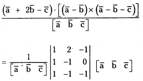
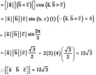
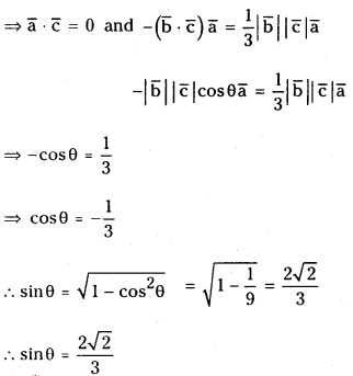
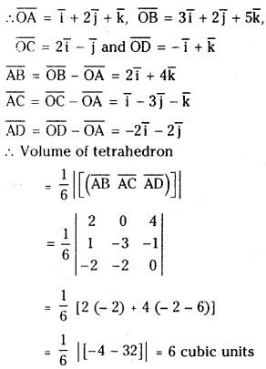
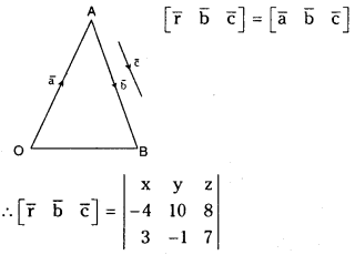
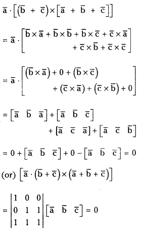
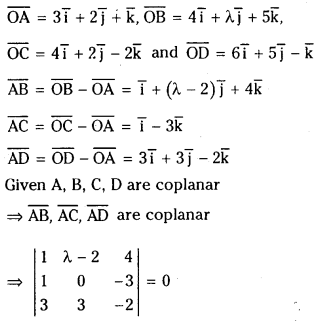
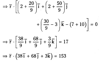
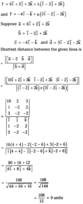
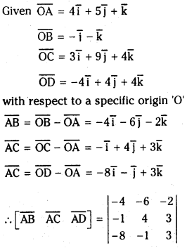
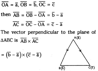


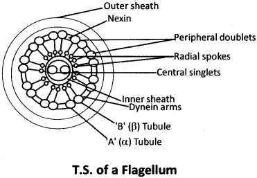
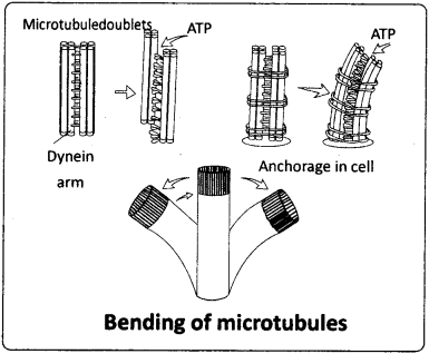
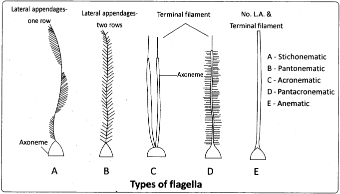
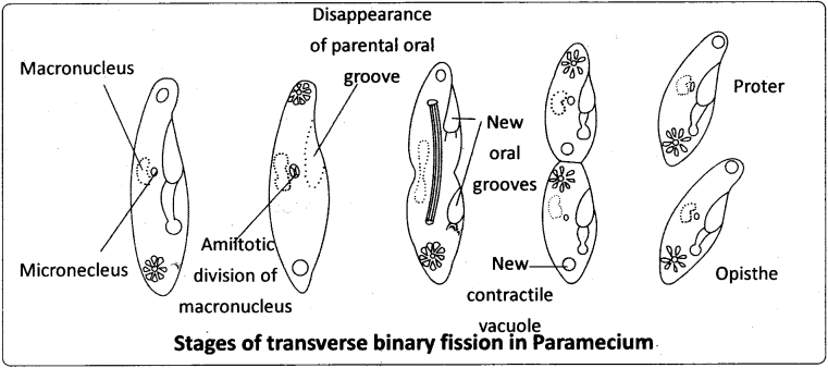
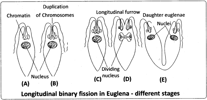
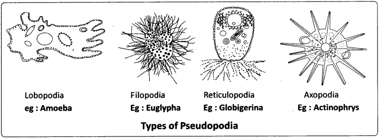
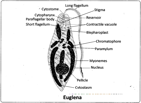
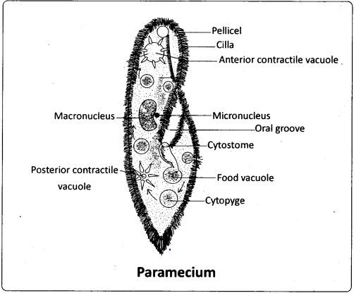
")
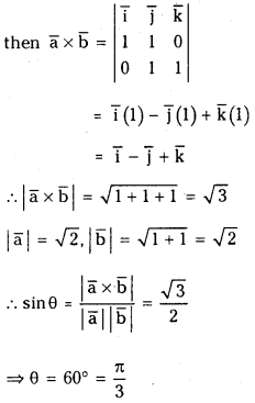


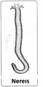
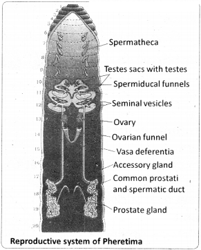

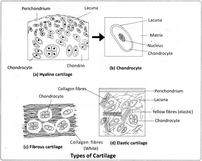
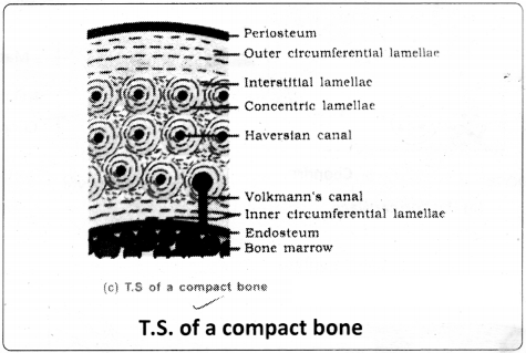
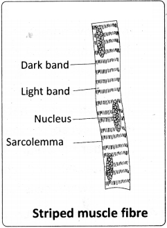
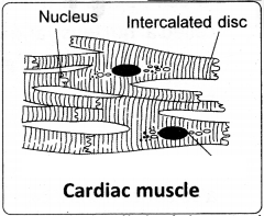
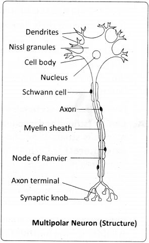
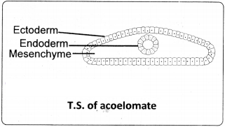
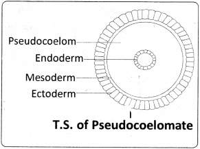
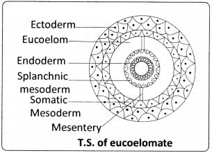
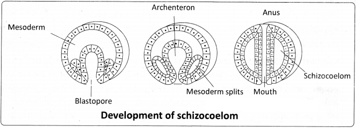
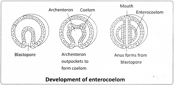
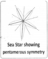
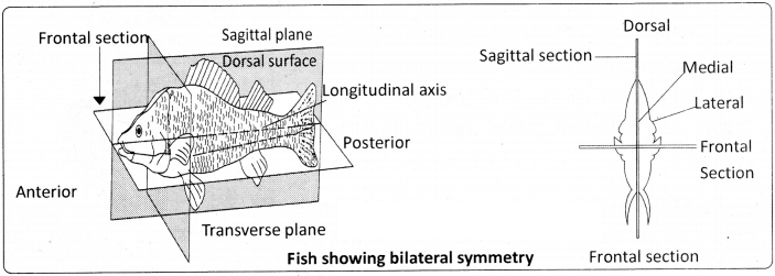
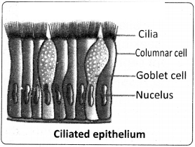
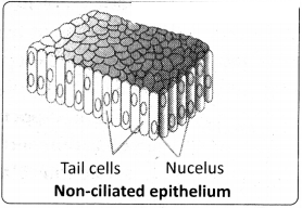
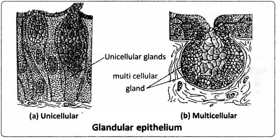
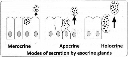
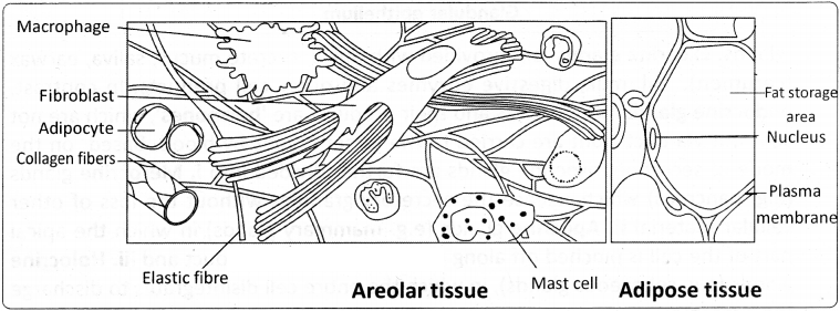
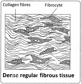
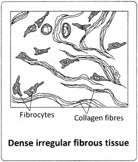
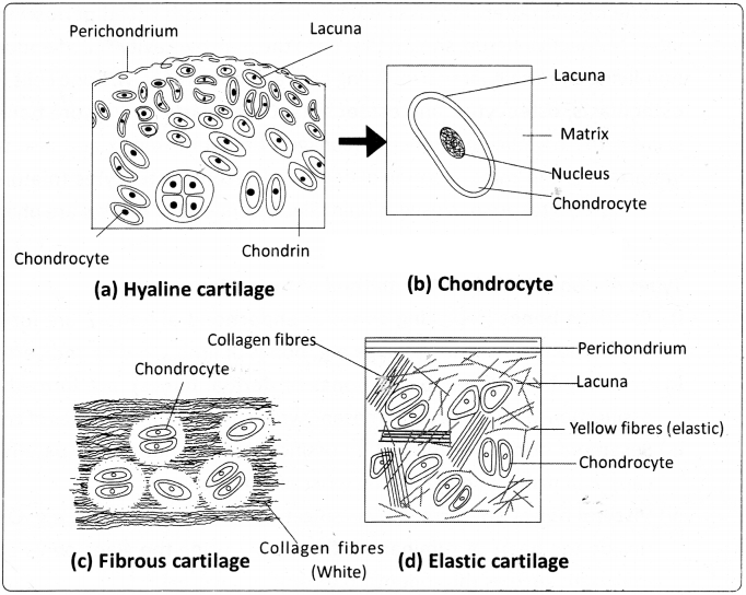
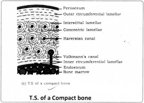
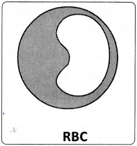
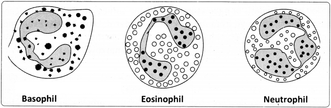
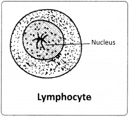
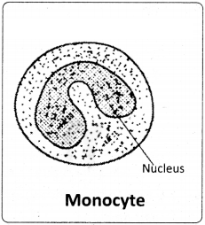

")